Application of bone morphogenetic protein-4 in drug for treating keratonosus

A corneal disease and protein technology, applied in the field of biopharmaceuticals, can solve problems such as aggravating corneal epithelial defects

Pending Publication Date: 2018-08-10
JILIN UNIV
View PDF2 Cites 0 Cited by
  • Summary
  • Abstract
  • Description
  • Claims
  • Application Information

AI Technical Summary

Problems solved by technology

The clinical application of epidermal growth factor and other drugs to promote the repair of corneal epithelial injury has achieved certain curative effect, but it has the risk of neovascularization and CNV.
But what is interesting is that currently the most effective anti-CNV treatment is bevacizumab, but its side effect is to aggravate corneal epithelial defect, which means that bevacizumab is not suitable for the same corneal epithelial defect CNV

Method used


Image

  • Application of bone morphogenetic protein-4 in drug for treating keratonosus
  • Application of bone morphogenetic protein-4 in drug for treating keratonosus
  • Application of bone morphogenetic protein-4 in drug for treating keratonosus

Examples


Embodiment 1

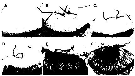
[0020] Example 1: Making a suture-induced rat CNV model, and observing the changes of CNV.

[0021] Animals were anesthetized with 8% chloral hydrate intraperitoneally before surgery. Tropicamide eye drops were applied topically (Santen, Osaka, Japan) with a 10-0 corneal suture (Johnson & Johnson Medical Ltd., St Stevens-Woluwe, Belgium) 1.5 mm from the corneoscleral limbus, through the epithelium and stroma layer, but did not penetrate the endothelial layer, were sutured two stitches. The distance between the two needles is 1mm. After operation, 0.3% ofloxacin eye ointment was applied to prevent infection. Over time, if the sutures are removed, the naturally existing natural blood vessels subside, therefore, the sutures remain until the end of treatment.

[0022] Such as figure 1 As shown, CNV growth was most active on day 7 after suture induction, forming a thick, dense capillary network. figure 1 Middle: A normal cornea, B 1 day after suture induction, corneal edema ne...

Embodiment 2

[0023] Example 2: Configuration and application of eye drops with BMP4 as active ingredient.

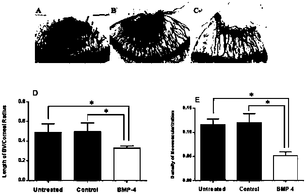
[0024] Get 4mM HCL to dissolve BMP4 and make the BMP4 solution of 20ug / ml, select this time point to instill the BMP4 solution of 20ug / ml to the outer eyes of the suture-induced rat CNV model prepared in Example 1, 1 drop / time, 3 times / day, for 7 consecutive days (the corneal sutures were retained during this process), with 4mM HCL solution as the control group. Such as figure 2 As shown, it was found that the area of ​​CNV in the dripping group was significantly smaller than that of the control group after 7 days of dripping. Such as image 3As shown, the ELISA kit was used to detect the protein levels of VEGF, TNFa, and MMP-9 in the cornea. A shows the ELISA results of corneal VEGF and BMP4. No differences were seen. B shows the ELISA results of corneal MMP-9 and TNF-α, and it was found that both of them were significantly decreased in the treatment group. The experimental gr...

Embodiment 3

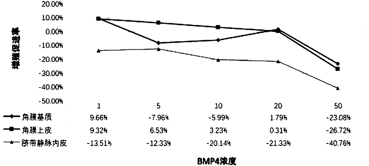
[0026] Example 3: To study the relationship between BMP4, VEGF and MMP-9 in the repair process of corneal injury.

[0027] MTT cell activity assay experiments were carried out on rat corneal epithelial cells, corneal stromal cells, and umbilical vein endothelial cells. A series of concentration gradients of BMP4 were designed to act on the three types of cells, and finally MTT was generated by lysing the cells succinate dehydrogenase. The formazan crystals were measured in a microplate reader to obtain experimental data. The epithelial scraping method was used to establish the corneal epithelial injury model, and the steps were as follows: Wistar rats were used as experimental animals, and the experiment was divided into normal tissue detection part and epithelial damage tissue detection part, paraffin sections were made, and TUNEL and HE staining were performed. To culture corneal epithelial cells, add BMP4 protein and VEGF antibody to the culture medium and set up solvent co...


PUM


Abstract

The invention belongs to the field of bio-pharmaceuticals, and particularly relates to application of bone morphogenetic protein-4 in a drug for treating keratonosus. The BMP4 has a mild growth-promoting effect on corneal epithelia and stromata, and treats CNV caused by epithelial damage; the BMP4 can serve as a drug for repairing damaged epithelia; the BMP4 is used for inhibiting keratitis reactions, and plays an important effect in the corneal injury repairing process, and the delivery way of t he BMP4 is convenient without trauma, and the BMP4 is easy to popularize.

Description

technical field [0001] The invention belongs to the field of biopharmaceuticals, and in particular relates to the use of bone morphogenic protein-4 in medicine for treating corneal diseases. Background technique [0002] Corneal disease is a common clinical disease and one of the most important blinding eye diseases in the world. Under normal circumstances, the cornea is an avascular, transparent connective tissue, which is the first barrier of the ocular surface and an important part of the refractive system. The cornea is exposed to the outside world and is easily affected by microorganisms, trauma, chemical and physical factors, and there are many chances of damage. Moreover, the cornea has no blood vessels and the supply of nutrients is limited. Therefore, once microorganisms invade, it is prone to infection. Among them, the corneal epithelium is located in the outermost layer of the cornea and is more vulnerable to attack by pathogenic microorganisms and the like. [...

Claims


Application Information

Patent Timeline
Patent Type & Authority Applications(China)
IPC IPC(8): A61K38/18A61P27/02
Inventor 张妍王淑荣何宇茜姚博远徐丽娜马岩
Owner JILIN UNIV
Why Eureka
Try Eureka