Antibody chip for detecting colorectal cancer related factors
An antibody chip, colorectal cancer technology, applied in measuring devices, biological testing, material inspection products, etc., can solve the problem of single detection index, achieve simple operation, fast detection, and save detection time
- Summary
- Abstract
- Description
- Claims
- Application Information
AI Technical Summary
Problems solved by technology
Method used
Image
Examples
Embodiment 1
[0048] The preparation of embodiment 1 antibody chip
[0049] (1) Antibody screening: Western blot experiments were used to identify the specificity and titer of the antibodies corresponding to the three preferred proteins, and to screen high-quality antibodies.
[0050] (2) Purified antibody: use Replace the antibody solvent with PBS in Ultra 10K ultrafiltration tubes, adjust the antibody concentration to 2mg / ml, aliquot 5ul, and store at -20°C.
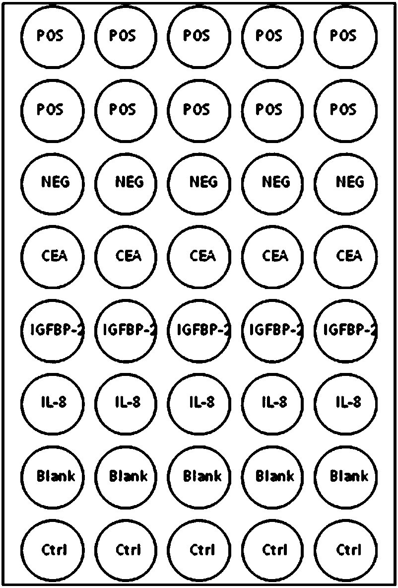
[0051] (3) Prepare spotting plate: add 18ul spotting buffer (PBS) to 2ul antibody, mix well, and add to spotting plate; add 18ul positive control (biotin-labeled secondary antibody) and 18ul negative control (PBS) to spotting plate .
[0052](4) Spotting: According to the operating requirements of the chip spotting instrument, do a good job of preparation before spotting, including lotion, system fluid, humidity, temperature, spotting needle, substrate and spotting plate, etc. After sample application, the chip was stored in a h...
Embodiment 2
[0054] Example 2 Application of antibody chip to detect multiple indicators in samples
[0055] 1. Sample Preparation
[0056] Let the venous blood stand for 20 minutes, centrifuge at room temperature at 3000r / min, separate the serum, and use it immediately; or divide it and store it in a -80°C refrigerator. Repeated freezing and thawing should be avoided. Thawed samples should be centrifuged again before testing.
[0057] 2. Biotin labeling of samples
[0058] (1) Sample centrifugation: centrifuge the sample at 4°C, 13,200 rpm for 10 minutes, and transfer the supernatant to a new centrifuge tube;
[0059] (2) Sample labeling: Take 3ul of each sample, put it into a centrifuge tube, add 2ul Biotin / DMF, and use the labeling buffer to make up the final volume of the reaction to 70ul. Mix well, shake and react at room temperature for 2h. Add 30ul of stop solution, shake at room temperature for 30min.
[0060] 3. Preparation of Standards
[0061] (1) Add 500ul sample diluent ...
Embodiment 3
[0080] Example 3 Sample Detection Specificity and Sensitivity Analysis
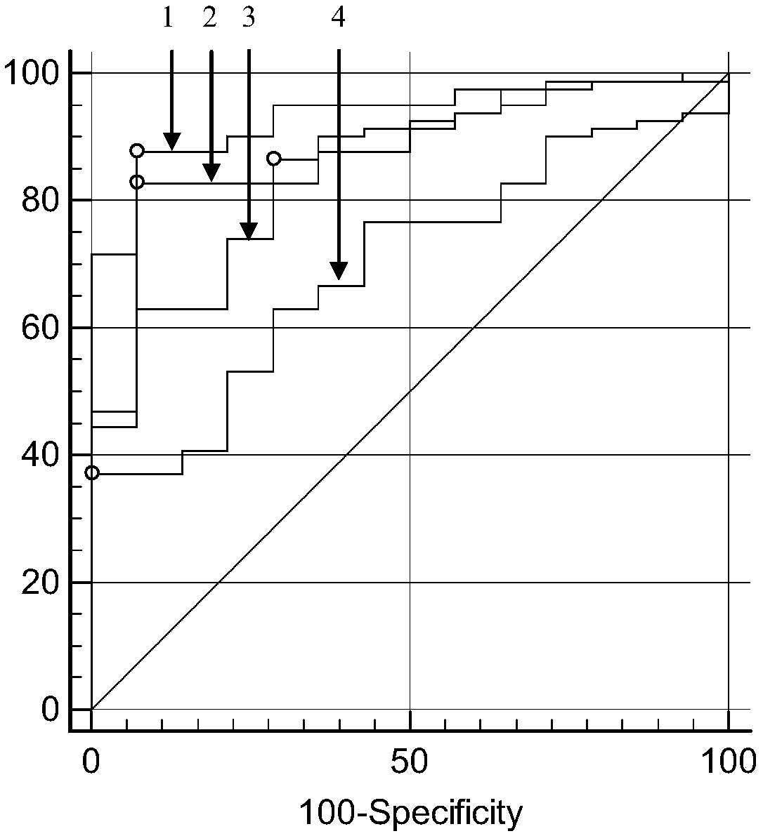
[0081] Using 95 cases of serum samples, the specificity and sensitivity of the kit of the present invention were tested. The detection cutoff value of CEA was 2479pg / ml, that of IGFBP-2 was 7785pg / ml, and that of IL-8 was 445pg / ml. Receiver operator characteristic curve (ROC) analysis was performed using Medcalc software to obtain data such as sensitivity, specificity and area under the curve (AUC). The sensitivity, specificity and AUC of each detection index to the detection of colorectal cancer serum samples are shown in Table 3. image 3 with Figure 4 .
[0082] Table 3 The sensitivity, specificity and AUC of each detection index to the detection results of colorectal cancer serum samples
[0083] Detection Indicator
[0084] It can be seen from the above table that the performance of the joint detection of the three indicators is better than that of each indicator alone.
PUM
| Property | Measurement | Unit |
|---|---|---|
| diameter | aaaaa | aaaaa |
Abstract
Description
Claims
Application Information
 - R&D
- Intellectual Property
- Life Sciences
- Materials
- Tech Scout
- Unparalleled Data Quality
- Higher Quality Content
- 60% Fewer Hallucinations
Browse by: Latest US Patents, China's latest patents, Technical Efficacy Thesaurus, Application Domain, Technology Topic, Popular Technical Reports.
© 2025 PatSnap. All rights reserved.Legal|Privacy policy|Modern Slavery Act Transparency Statement|Sitemap|About US| Contact US: help@patsnap.com


