Oral patch preparing method of composite oral mucosa epithelial cells

A technology of oral epithelial cells and oral mucosa, applied in the field of cell biology, can solve the problems of difficulty in culturing oral epithelial cells, difficulty in obtaining oral prosthetic epithelial cells, etc. Effect

Inactive Publication Date: 2018-09-07
上海白衣缘生物工程有限公司
View PDF9 Cites 0 Cited by
  • Summary
  • Abstract
  • Description
  • Claims
  • Application Information

AI Technical Summary

Problems solved by technology

[0007] The technical problems solved by the present invention are: 1) It is difficult to culture oral epithelial cells, and it is difficult to obtain the amount of epithelial cells for oral restoration surgery

Method used


Image

  • Oral patch preparing method of composite oral mucosa epithelial cells
  • Oral patch preparing method of composite oral mucosa epithelial cells

Examples


Embodiment 1

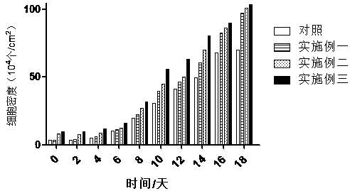
[0036] 1. Preparation of oral patch: The oral patch was prepared using the submucosa. The steps are to obtain the material from the submucosa, and obtain it after decellularization, DNA removal, virus inactivation, freeze-drying and irradiation sterilization;

[0037]2. Primary culture of oral mucosal epithelial cells: Obtain oral mucosal tissue in the oral cavity of surgical patients under sterile conditions. Rinse repeatedly with double-antibiotic saline containing 100ug / ml penicillin and 100ug / ml streptomycin, then rinse with PBS solution, remove submucosal tissue with ophthalmic scissors, cut into small pieces of about 5mm×5mm, and put them into a petri dish Inside, add DKSFM medium containing 0.25% Dispase II and submerge the tissue block, digest at 4°C for 16 hours, and separate the superficial epithelium and subepithelial layer with ophthalmic forceps;

[0038] The epithelial layer was digested with 0.25% trypsin at 37°C for 15 minutes (5 min, pipetting and vibration o...

Embodiment 2

[0046] 1. Preparation of oral patch: The oral patch was prepared using the submucosa. The steps are to obtain the material from the submucosa, and obtain it after decellularization, DNA removal, virus inactivation, freeze-drying and irradiation sterilization;

[0047] 2. Primary culture of oral mucosal epithelial cells: Obtain oral mucosal tissue in the oral cavity of surgical patients under sterile conditions. Rinse repeatedly with double-antibiotic saline containing 100ug / ml penicillin and 100ug / ml streptomycin, then rinse with PBS solution, remove submucosal tissue with ophthalmic scissors, cut into small pieces of about 5mm×5mm, and put them into a petri dish Inside, add DKSFM medium containing 0.25% Dispase II and submerge the tissue block, digest at 4°C for 17 hours, and separate the superficial epithelium and subepithelial layer with ophthalmic forceps;

[0048] The epithelial layer was digested with 0.25% trypsin at 37°C for 15 minutes (5 min, pipetting and vibration ...

Embodiment 3

[0056] 1. Preparation of oral patch: The oral patch was prepared using the submucosa. The steps are to obtain the material from the submucosa, and obtain it after decellularization, DNA removal, virus inactivation, freeze-drying and irradiation sterilization;

[0057] 2. Primary culture of oral mucosal epithelial cells: Obtain oral mucosal tissue in the oral cavity of surgical patients under sterile conditions. Rinse repeatedly with double-antibiotic saline containing 100ug / ml penicillin and 100ug / ml streptomycin, then rinse with PBS solution, remove submucosal tissue with ophthalmic scissors, cut into small pieces of about 5mm×5mm, and put them into a petri dish Inside, add DKSFM medium containing 0.25% Dispase II and submerge the tissue block, digest at 4°C for 18 hours, and separate the superficial epithelium and subepithelial layer with ophthalmic forceps;

[0058] The epithelial layer was digested with 0.25% trypsin at 37°C for 15 minutes (5 min, pipetting and vibration ...


PUM


Abstract

The invention discloses an oral patch preparing method of composite oral mucosa epithelial cells, which includes the following steps: preparing the oral patch by using submucosa; culturing the oral mucosa epithelial cells by adopting a subculture method; inoculating the oral mucosa epithelial cells into a cell culture plate with the oral patch; culturing in a cell incubator with the temperature of37 DEG C and 5% CO2, and the oral patch of the composite oral mucosa epithelial cells is obtained once the culture is finished. The oral patch preparing method of the composite oral mucosa epithelialcells has the advantage that material sources are adequate, the oral mucosa epithelial cells can largely proliferate in the oral patch, and a new way is provided for clinic reconstruction oral mucosalarge area defects.

Description

technical field [0001] The invention relates to a method for preparing an oral patch made of composite oral mucosa epithelial cells, belonging to the field of cell biology technology. Background technique [0002] Oral mucosa, as the first line of defense of the human body's biological barrier, plays a vital role in daily life, but the location of the oral mucosa and its surrounding environment lead to a variety of factors that can cause loss of oral mucosal tissue, such as oral and maxillofacial surgery Tumor resection, trauma, and alveolar surgery before repair often have large oral mucosal defects, which are often repaired clinically by skin grafting or flap transfer, but skin grafting often causes oral cavity narrowing and affects function due to contracture, and local flaps Metastasis often causes local swelling and poor shape, which is difficult for patients to accept. Moreover, autologous tissue transplantation is a treatment mode of "repairing damage by trauma" at t...

Claims


Application Information

Patent Timeline
Patent Type & Authority Applications(China)
IPC IPC(8): A61L27/36A61L27/38
CPCA61L27/3604A61L27/3629A61L27/3641A61L27/3666A61L27/3687A61L27/3691A61L27/3813A61L27/3839A61L27/3869A61L27/3895A61L27/50A61L2300/412A61L2430/40C12N5/0632C12N2509/00
Inventor 李康乐汪文涛葛翠兰
Owner 上海白衣缘生物工程有限公司
Why Eureka
Try Eureka