Multilayer bionic joint based on curved surface 3D printing and preparation method thereof

A bionic joint and 3D printing technology, applied in 3D printing, joint implants, joint implants, etc., can solve problems that cannot meet joint repair, cannot simulate calcified layers well, and are difficult to achieve cartilage microenvironment and osteogenesis Issues such as effective isolation of the microenvironment

Pending Publication Date: 2020-12-15
AFFILIATED ZHONGSHAN HOSPITAL OF DALIAN UNIV +1
View PDF4 Cites 4 Cited by
  • Summary
  • Abstract
  • Description
  • Claims
  • Application Information

AI Technical Summary

Problems solved by technology

CN105105875A designed a bionic artificial hip joint with in-growth function, but its articular surface wrapping angle is only 60-120°, which cannot meet the repair of joints when a large area of ​​osteochondral joint is damaged

Method used


Image

  • Multilayer bionic joint based on curved surface 3D printing and preparation method thereof
  • Multilayer bionic joint based on curved surface 3D printing and preparation method thereof
  • Multilayer bionic joint based on curved surface 3D printing and preparation method thereof

Examples


Embodiment 1

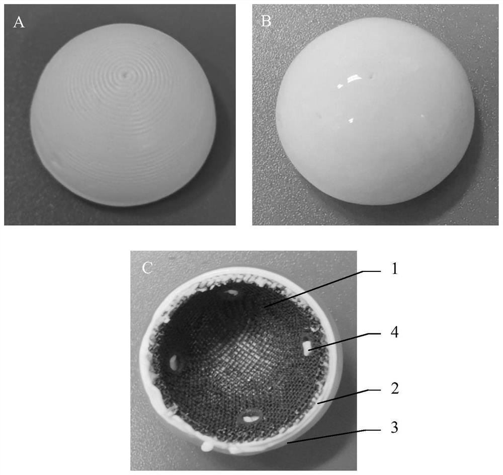
[0058] A multi-layer bionic joint, the multi-layer bionic joint is closely contacted according to the order of the inner layer, the middle layer and the outer layer, the inner layer is a porous tantalum metal support, the middle layer is a solid support, and the outer The layer is a solid gelatin / sodium alginate composite hydrogel scaffold, and the inner layer, the middle layer and the outer layer all have an arc-shaped shell structure, and the arc-shaped shell is a hemispherical shell.

[0059] The inner layer is made of tantalum metal powder by metal 3D printing, and the middle layer is deposited on the inner layer with hydroxyapatite slurry as the base material by bio-3D printing. The outer layer is made by depositing gelatin / sodium alginate composite hydrogel liquid as a base material on the outer surface of the middle layer by bio-3D printing. The inner surface of the intermediate layer is in close contact with the outer surface of the inner layer in a manner of covering ...

Embodiment 2

[0079] A multilayer bionic joint with a cell loading cavity is prepared by the following method:

[0080] (1) A three-layer 3D printed bionic joint with a three-layer structure is prepared through steps 1) to 8) described in Example 1;

[0081] (2) The three-layer 3D printed bionic joint cross-linked in 5% calcium chloride solution for 10 minutes was put into a shaped mold to form a cell loading cavity on the outer surface.

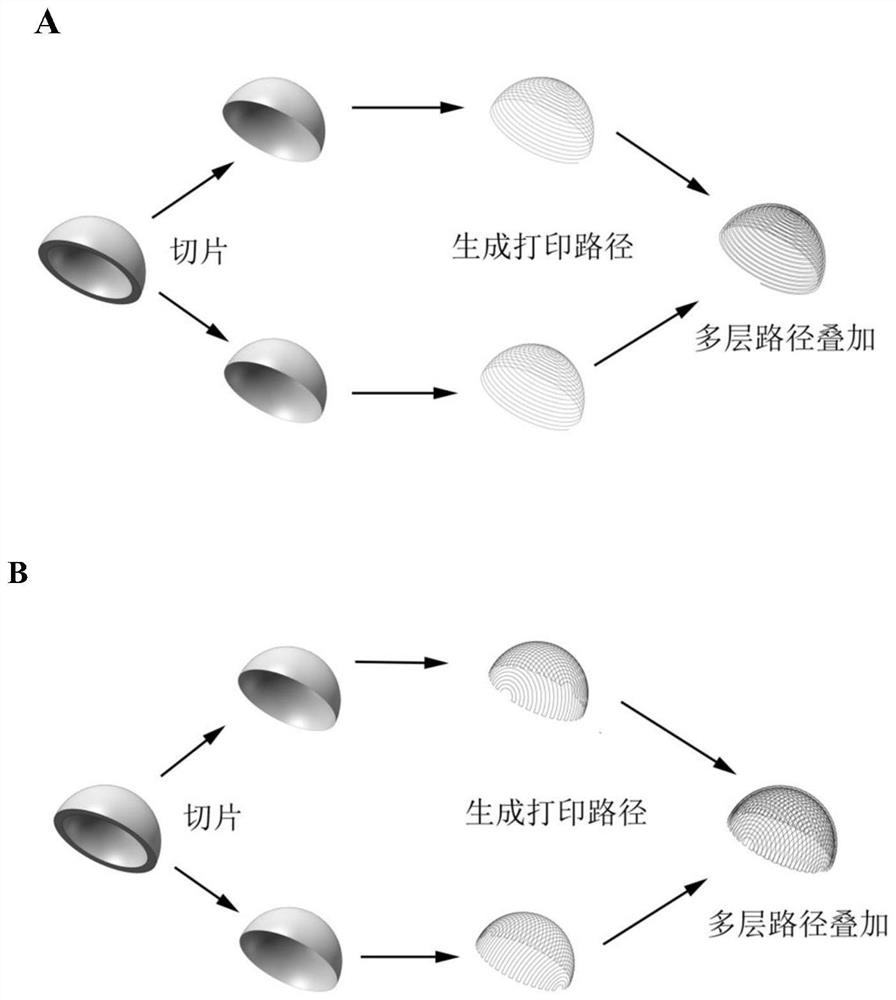
[0082] Figure 5 is a structural schematic diagram of a shaped mold for forming a cell loading cavity, wherein, Figure 5 A to C are the schematic diagram, top view and side view of the three-dimensional geometric model designed with drawing software, respectively. Figure 5 D is the physical map of the titanium alloy sizing mold after 3D printing based on the three-dimensional geometric model. When the software designs the sizing mold, its size and shape are the same as the three-dimensional geometric model of the bionic joint of the present invention (...

Embodiment 3

[0085] The preparation method of the multilayer bionic joint based on 3D printing comprises the following steps:

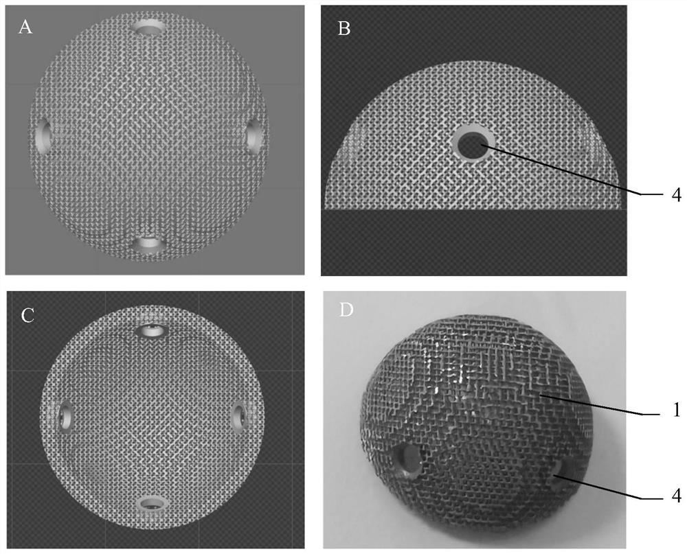
[0086] 1) According to the method described in steps 1) to 2) in Example 1, the porous tantalum metal powder is used as the matrix, and the porous tantalum metal hemispherical shell (such as figure 1 );

[0087] Among them, the 3D printing conditions are: the thickness of powder coating is 30μm, the laser power is 200W, the exposure time is 70μs, the laser scanning point spacing is 40μm, and the line spacing is 40μm; wherein, the porous tantalum metal powder is a spherical powder of medical grade, and its purity is ≥ 99.99% by weight, particle size 30 μm, the protective gas is high-purity argon (purity ≥ 99.99%), and the oxygen content in the working chamber is less than 1000ppm during the printing process;

[0088] 2) The printed parts are subjected to high-temperature heat treatment to eliminate residual stress generated during processing, and to smooth the su...


PUM

PropertyMeasurementUnit
Radianaaaaaaaaaa
Widthaaaaaaaaaa
The inside diameter ofaaaaaaaaaa

Abstract

The invention discloses a bionic bone joint based on curved surface 3D printing and a preparation method thereof. The multilayer bionic joint is formed by an inner layer, a middle layer and an outer layer which are in close contact in sequence, wherein the inner layer is a porous tantalum metal support, the middle layer is a solid biological ceramic support, the outer layer is a solid gelatin/ sodium alginate composite hydrogel support, the inner layer, the middle layer and the outer layer are all of an arc-shaped shell structure, the radian of the arc-shaped shell is 120-240 degrees, and a cell loading cavity formed by continuous or discontinuous edges formed by protruding outwards from the surface of the outer layer and grooves formed between the edges is formed in the surface of the outer layer. The multilayer bionic joint is large in arc surface radian and suitable for repairing large-area osteochondral joint defects, and the repairing area can be larger than 1/2 of the area of thewhole joint. Cells are inoculated into the cell loading cavity, so that the adhesion rate of the cells is increased, and the problem that the surface of the outer layer of the bionic joint is smooth,so that the inoculated cells are not prone to adhesion is solved.

Description

technical field [0001] The invention relates to the technical field of medical osteochondral repair, in particular to a bionic bone joint based on curved surface 3D printing and a preparation method thereof. Background technique [0002] Osteochondral joint disease is a common disease in orthopedics, and there is a lack of effective treatment methods. The main methods used in the clinical repair of osteochondral defects include debridement, bone marrow stimulation, osteochondral grafting, and joint replacement. For small-area injuries, osteochondral transplantation is currently the main clinical treatment method, but there are problems such as the limitation of the amount of material taken, damage to the donor site, and difficulty in matching the graft with the articular surface; for large-area osteochondral joint defects, the most clinically A common method is joint replacement. However, the artificial joint prosthesis has complications such as infection, loosening, and s...

Claims


Application Information

Patent Timeline
IPC IPC(8): A61F2/30
CPCA61F2/30756A61F2002/3006A61F2002/30948A61F2002/30985
Inventor 赵德伟尉晓蔚李军雷刘阁吴斌徐铭恩
Owner AFFILIATED ZHONGSHAN HOSPITAL OF DALIAN UNIV
Why Eureka
Try Eureka