

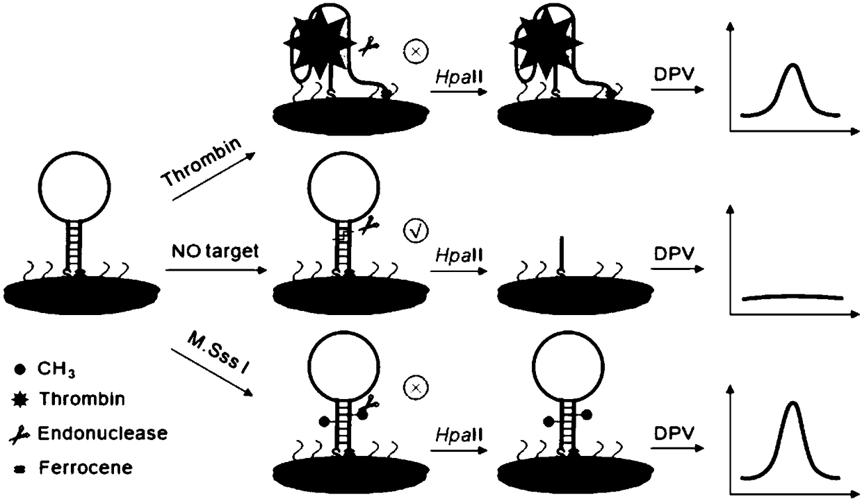
Aptamer probe for detecting two kinds of tumor makers and electrochemical biosensor and preparation method and application thereof
A technology of tumor markers and biosensors, applied in the direction of material electrochemical variables, scientific instruments, instruments, etc., can solve problems affecting the analytical performance of sensors
- Summary
- Abstract
- Description
- Claims
- Application Information
AI Technical Summary
Problems solved by technology
Method used
Image

Examples
Embodiment 1
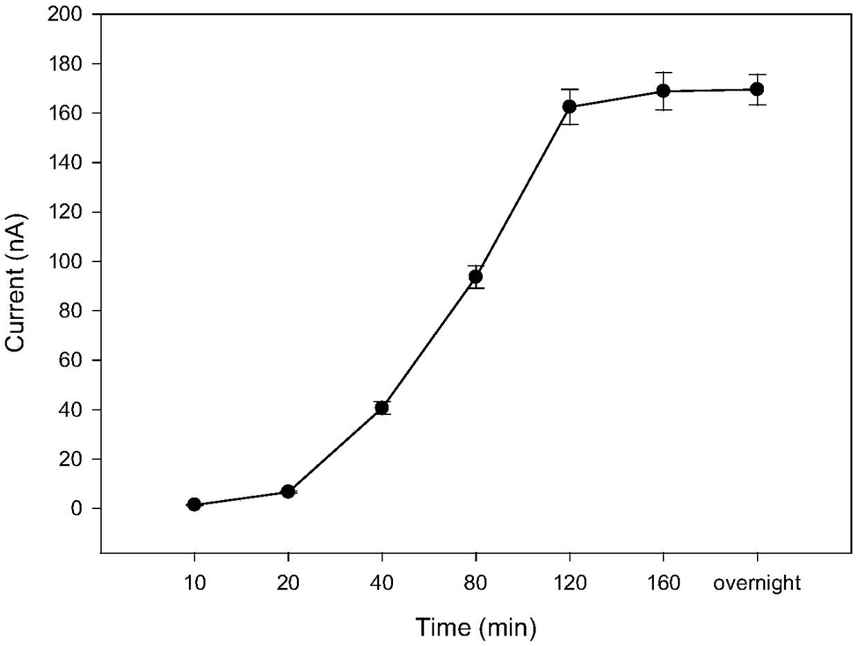
[0055] Example 1 Optimization of assembly time of the aptamer probe of the present invention
[0056] In this embodiment, the method for optimizing the assembly time of the aptamer probe includes the following steps:
[0057] (1) Polish the gold electrode on the suede in 0.3 μm and 0.05 μm alumina slurry until it becomes a mirror surface, rinse with distilled water, and then ultrasonicate in secondary water, ethanol, and secondary water for 5 minutes, and then Soak the electrode in Piranha solution for 15min, rinse with secondary water and place the gold electrode in 0.1M H 2 SO 4 The cyclic voltammetry scan was performed between -0.3V and +1.5V, and then the cleaned gold electrode was rinsed with secondary water.
[0058] (2) 20 μL of 3.8 μM aptamer probe was dropped onto the surface of the pretreated gold electrode, and self-assembled at room temperature for different times (10 min, 20 min, 40 min, 80 min, 120 min, 160 min, overnight).
[0059] (3) After the gold electrod...
Embodiment 2
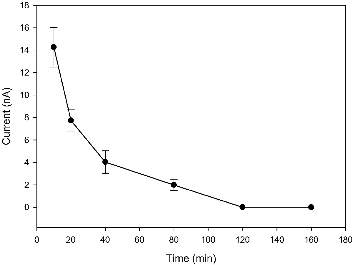
[0062] Embodiment 2 Hpa II endonuclease digestion time optimization
[0063] In the present embodiment, the Hpa II endonuclease digestion time optimization method comprises the following steps:
[0064] (1) Polish the gold electrode on the suede in 0.3 μm and 0.05 μm alumina slurry until it becomes a mirror surface, rinse with distilled water, and then ultrasonicate in secondary water, ethanol, and secondary water for 5 minutes, and then Soak the electrode in Piranha solution for 15min, rinse with secondary water and place the gold electrode in 0.1M H 2 SO 4 The cyclic voltammetry scan was performed between -0.3V and +1.5V, and then the cleaned gold electrode was rinsed with secondary water.
[0065] (2) 20 μL of 3.8 μM aptamer probe was dropped onto the surface of the pretreated gold electrode, and self-assembled at room temperature for 2 h.
[0066] (3) After the gold electrode was washed with secondary water, 20 μL of 1 mM mercaptohexanol was dropped onto the surface of ...
Embodiment 3
[0070] Embodiment 3Hpa II endonuclease concentration optimization
[0071] In the present embodiment, the Hpa II endonuclease concentration optimization method comprises the following steps:
[0072] (1) Polish the gold electrode on the suede in 0.3 μm and 0.05 μm alumina slurry until it becomes a mirror surface, rinse with distilled water, and then ultrasonicate in secondary water, ethanol, and secondary water for 5 minutes, and then Soak the gold electrode in Piranha solution for 15min, rinse with secondary water and put the gold electrode in 0.1M H 2 SO 4 The cyclic voltammetry scan was performed between -0.3V and +1.5V, and then the cleaned gold electrode was rinsed with secondary water.
[0073] (2) 20 μL of 3.8 μM aptamer probe was dropped onto the surface of the pretreated gold electrode, and self-assembled at room temperature for 2 h.
[0074] (3) After the gold electrode was washed with secondary water, 20 μL of 1 mM mercaptohexanol was dropped onto the surface of ...
PUM
 Abstract
Description
Claims
Application Information