Preparation method of microneedle array biosensor

A technology of biosensors and microneedle arrays, applied in sensors, medical science, blood characterization devices, etc., can solve the problems of polymer microneedle arrays not being strong enough, easily causing infection, and wounds that cannot heal within minutes. Realize the effect of maximum utilization rate and simple preparation process

Pending Publication Date: 2018-11-20
SUN YAT SEN UNIV
View PDF7 Cites 2 Cited by
  • Summary
  • Abstract
  • Description
  • Claims
  • Application Information

AI Technical Summary

Problems solved by technology

For example, for blood glucose detection, which is common in in vivo detection, the use of microneedle array biosensors for blood glucose detection does not require blood collection, and can directly measure the glucose content in interstitial fluid. The glucose content in interstitial fluid is basically the same as the blood glucose content in blood. There is a delay of 5-10 minutes, and if the common blood glucose meter on the market is used for blood sugar testing, patients need to measure blood sugar anytime and anywhere by fingertip blood, but fingertip blood will cause pain, and fingertip blood Bloody wounds cannot heal within minutes and are prone to infection
[0004] Although microneedle array biosensors can be used for in vivo detection, the materials traditionally used to make microneedle arrays are mainly metals, single crystal silicon and polymers, and single crystal silicon microneedle arrays are relatively brittle, causing the microneedles to easily break
Polymer microneedle arrays are not strong enough to pierce the stratum corneum. Metal microneedle arrays are relatively mature and safe, but these types of microneedle arrays all have a common shortcoming, that is, the prepared microneedle arrays The arrays are all smooth surfaces, and the biorecognition molecules modified on the surface of the microneedle array will fall off due to friction during the puncture process, causing the microneedle array entering the body to fail to achieve the purpose of in vivo detection

Method used


Image

  • Preparation method of microneedle array biosensor
  • Preparation method of microneedle array biosensor

Examples


Embodiment 1

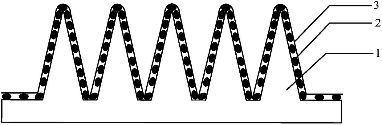
[0022] like figure 1 As shown, the microneedle array biosensor includes a microneedle array 1, a biorecognition molecule 2 modified on the surface of the microneedle array 1, and a sprayed degradable polymer material 3. The microneedle array biosensor in Example 1 is used for glucose in body detection. The specific preparation method is as follows:

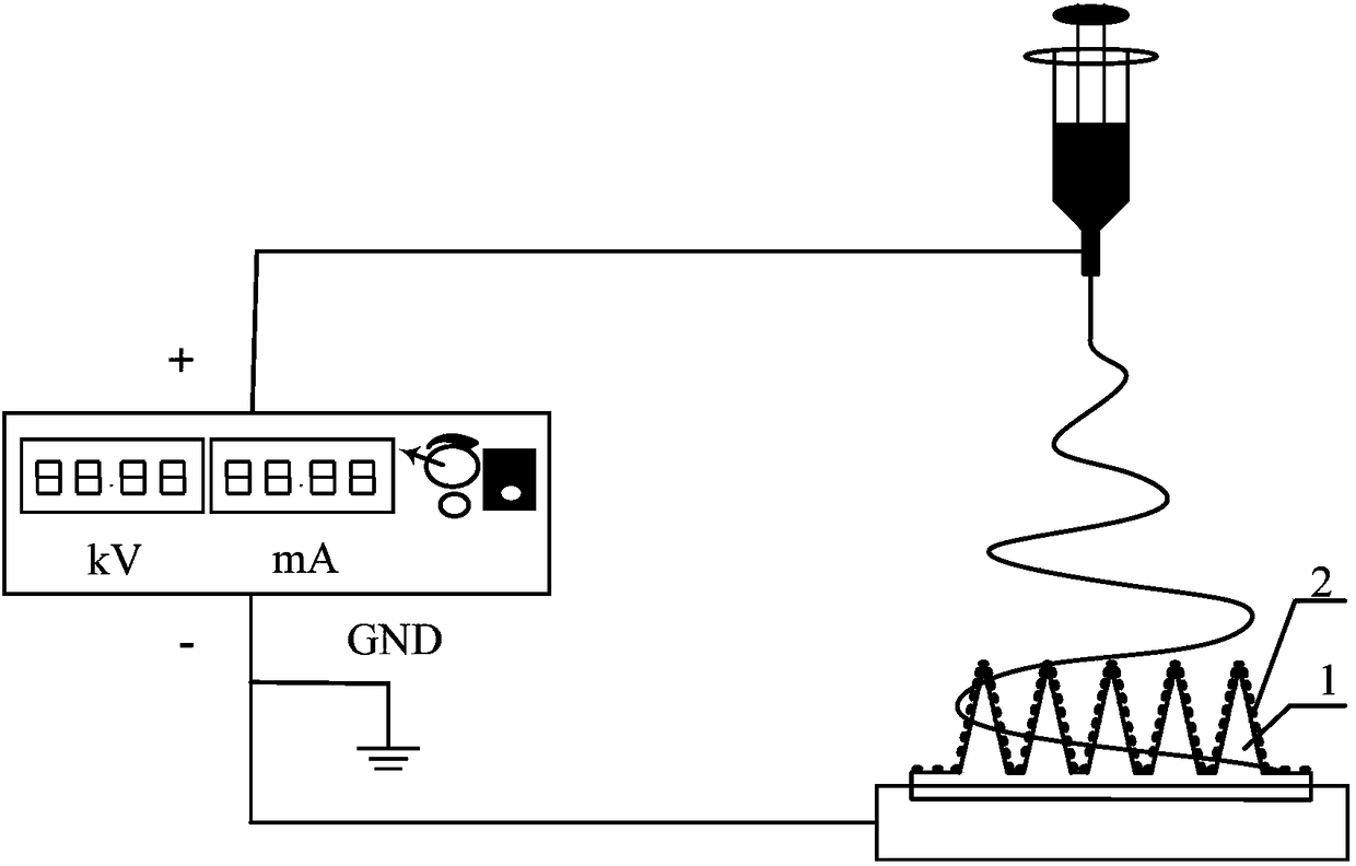
[0023] a. Preparation of microneedle array 1: Laser drilling is used to prepare a microneedle array mold, and a gradient rough surface is formed on the inner cavity of the microneedle array mold during the process of preparing the microneedle array mold; the liquefied, conductive The microneedle array material is poured in the microneedle array mould, and demolded to form a conductive microneedle array, the material of the microneedle array mold is an aluminum sheet, the laser drilling speed is set to 200mm / s, and the number of times of drilling is set to At 1200 times, under the above setting conditions, the aluminum sheet perf...

Embodiment 2

[0028] The difference between this embodiment 2 and the foregoing embodiment 1 is that the microneedle array biosensor of this embodiment 2 is used for in vivo detection of cholesterol. The enzyme molecule selected in step b is 50 uL of cholesterol oxidase at 10 mg / ml. Like Example 1, the enzyme molecule is also modified on the surface of the microneedle array 1 by a chemical cross-linking method, and the selected chemical cross-linking agent is still glutaraldehyde. The choice of 10mg / ml cholesterol oxidase is the optimal concentration obtained after parameter optimization of its concentration, and at this concentration, the microneedle array biosensor has the maximum response. The selected concentration of glutaraldehyde is 0.2%, soaked in the environment of 4 ℃ for 4 hours, and cross-linking for 4 hours is the best time for experimental optimization. When cross-linking for 4 hours, cholesterol oxidase can be immobilized on the microneedle array organism with maximum efficie...


PUM


Abstract

The invention discloses a preparation method of a microneedle array biosensor. The preparation method of the microneedle array biosensor comprises the following steps: making a conductive microneedlearray with rough surface; modifying a biological recognition molecule on the rough surface of the microneedle array; adding a degradable macromolecular material on the surface, on which the biologicalrecognition molecule is modified, of the microneedle array. The microneedle array in the microneedle array biosensor has the rough surface, so that protection of the biological recognition molecule modified on the surface of the microneedle array in the puncture process is benefited; furthermore, the degradable micromolecular material added on the outer layer of the microneedle array can realizesecondary protection of the biological recognition molecule.

Description

technical field [0001] The invention relates to the field of biomedical engineering, in particular to a method for preparing a microneedle array biosensor. Background technique [0002] The microneedle array biosensor is a biorecognition molecule modified on the surface of the microneedle array. In the in vivo detection, after the microneedle array biosensor punctures into the skin, the biorecognition molecule directly contacts with the body fluid, and the biorecognition molecule will specifically interact with the tissue fluid. The biomolecules are specifically combined, and electrons are generated at the same time, and the concentration of biomolecules is expressed in the form of electrical signals. [0003] The use of microneedle array biosensors for in vivo detection has the advantages of little damage to human skin, no pain or slight pain, skin can be restored to its original state in a short time, and microneedle arrays inserted into the human dermis will not cause bio...

Claims


Application Information

Patent Timeline
Patent Type & Authority Applications(China)
IPC IPC(8): A61B5/145
CPCA61B5/14503A61B5/14532A61B5/685
Inventor 蒋乐伦高洁易长青
Owner SUN YAT SEN UNIV
Why Eureka
Try Eureka