Method of preparing cell lysate

a cell lysate and cell technology, applied in the field of cell lysate preparation, can solve the problems of time-consuming and laborious rt-pcr process, etc., and achieve the effects of simplifying the preparation of cell lysate, stabilizing the yield of recovered cytosolic rna, and facilitating fluctuation of recovered mrna

Inactive Publication Date: 2007-08-21
RESONAC CORPORATION +1
View PDF28 Cites 87 Cited by
  • Summary
  • Abstract
  • Description
  • Claims
  • Application Information

AI Technical Summary

Benefits of technology

[0008]Further, conventionally, cell lysate is prepared by vigorous homogenization processes which are not only time-consuming and labor-intensive, but also cause fluctuation of the amount of recovered mRNA. In the present invention, in combination with the PCR microplates, by placing target cells on a filter plane evenly and passing a lysis buffer through the cell layer on the filter without disturbing the cells, it is possible to drastically simplify the preparation of cell lysate and significantly stabilize the yield of recovered cytosolic RNA. It is very convenient if the microplate and the filter plate are available as a kit for direct RT-PCR. In the above, a lysis buffer, wash buffer, reagents for RT-PCR / PCR, and PBS are commercially available or can readily be prepared so that they need not be included in the kit. However, for convenience, a lysis buffer may be included in the kit for releasing cytosolic mRNA present in the cells when passing through the cell layer on the filter plate. In the above, the lysis buffer comprises a mild detergent for destructing cell membranes but maintaining nuclei to be intact and a reagent for inhibiting RNase activity or inactivating RNase, said lysis buffer having a pH and salt concentration for hybridization.
[0009]Because of the above features, the PCR microplates and filter plates can drastically and surprisingly simplify the entire process of RT-PCR from the preparation of cell lysate to measuring specific PCR products with high reliability. Thus, this technology is highly useful in various molecular analyses including basic research, diagnostics, and drug screening, with potential application to future automation.

Problems solved by technology

Heretofore, microplates were never used for PCR due to insufficient thermal stability, and thus, RT-PCR processes were time-consuming and labor-intensive.
Further, conventionally, cell lysate is prepared by vigorous homogenization processes which are not only time-consuming and labor-intensive, but also cause fluctuation of the amount of recovered mRNA.

Method used


Image

  • Method of preparing cell lysate
  • Method of preparing cell lysate
  • Method of preparing cell lysate

Examples


examples

[0050]The invention will be further explained with reference to Examples shown below. Materials and methods used in the Examples are as follows:

[0051]Materials: Oligo(dT)-immobilized oligonucleotide-immobilized PCR microplates (GENEPLATE®-PP, Hitachi Chemical Research Center, Irvine, Calif.), YOYO™-1 (1,1′-(4,4,7,7-tetramethyl-4,7-diazaundecamethylene)-bis-4-[3-methl-2,3-dihydro-(benzo-1,3-oxazole)-2-methylidene]-quinoliumetraiodide, Molecular Probes, Eugene, Oreg.), reagents for PCR (Taq polymerase, EZ rTth RNA-PCR kit) (Perkin Elmar, Foster City, Calif.), K562 cell line (American Type Culture Collection, Rockville, Md.), 100 bp DNA ladder, phosphate buffered saline (PBS), vanadyl ribonucleoside complex (VRC), rabbit globin mRNA, cell culture medium and appropriate antibiotics, buffer-saturated phenol (Gibco-BRL, Geithersburg, Md.), fetal bovine serum (FBS, HyClone, Logan, Utah), biotin-dUTP (Clontech, Palo Alto, Calif.), ATTOPHOS™ (alkaline phosphatase substrate, JBL Scientific, S...

experiment 1

of Immobilized Oligonucleotides

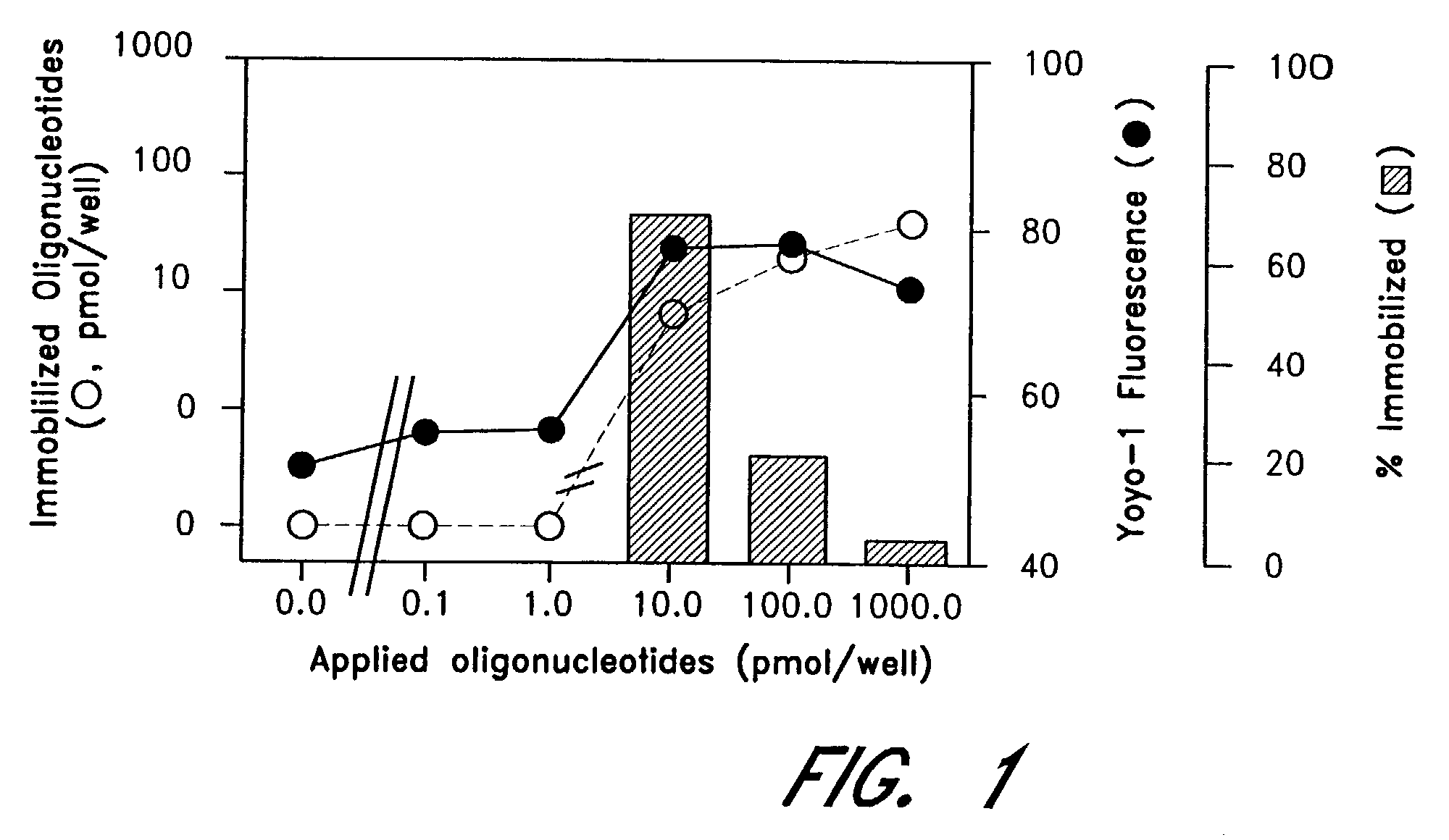
[0059]GENUNC™ PP microplates (Nunc, Naparville, Ill.) treated at Hitachi Chemical Research Center, Irvine, Calif., were obtained from AGCT, Irvine, Calif., and oligonucleotides were immobilized thereonto. Oligonucleotide concentrations were determined before and after immobilization as 1.0 OD260 unit equals to 30 μg / ml, and the amounts of immobilized oligonucleotides were calculated by subtracting one value from another. In separate experiments, YOYO-1 was diluted in TE (10 mmol / L Tris, pH 8.0, 1 mmol / L EDTA) in a final dilution of 1:1000, and applied to GENEPLATE®-PP microplates. The fluorescence was determined by CYTOFLUOR™ 2300 (Millipore, Bedford, Mass.) with excitation and emission wavelengths of 485 nm (bandwidth 20 nm) and 530 nm (band width 25 nm), respectively, as previously described (Miura Y, at al., Clin Chem 1996:42:1758-64, Miura Y, et al., Cancer Lett 1997:116:139-44).

[0060]FIG. 1 is a graph showing quantities of oligo(dT) immobilized on...

experiment 2

city of Oligonucleotide-Immobilized PCR Microplates

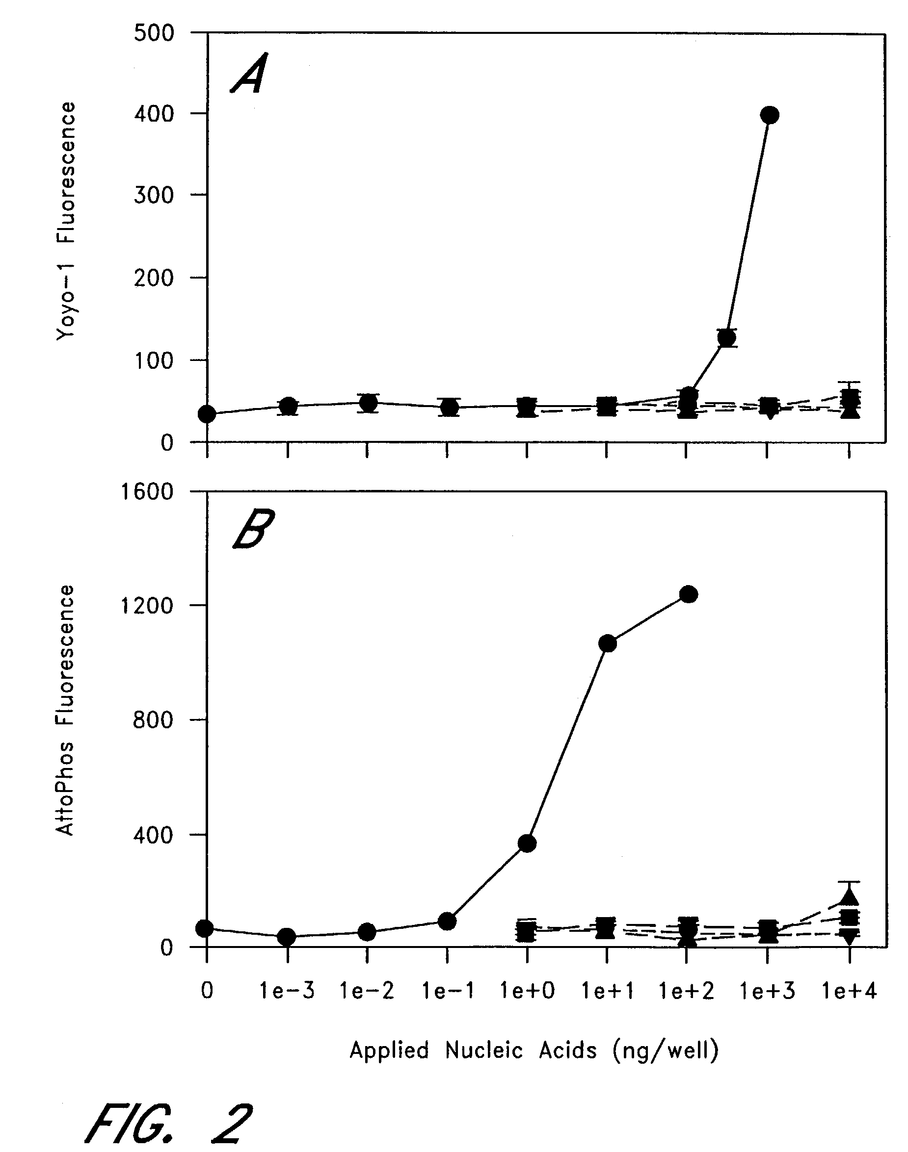
[0061]The next series of experiments was conducted to show mRNA specificity. FIG. 2A is a graph showing mRNA specificity of the oligonucleotide-immobilized PCR microplates, wherein YOYO-1 fluorescent intensity indicates high specificity to mRNA (●) over DNA (▪), rRNA (Δ), and tRNA (∇). FIG. 2B is a graph showing mRNA specificity of the oligonucleotide-immobilized PCR microplates, wherein substrate ATTOPHOS™ indicates high specificity to mRNA (●) over DNA (▪), rRNA (Δ), and tRNA (∇).

[0062]In the figures, various concentrations of rabbit globin mRNA (●), DNA (▪), rRNA (Δ), tRNA (∇) were suspended in 50 μl of hybridization buffer (10 mmol / L Tris, pH 8.0, 1 mmol / L EDTA, 0.5 M NaCl) and applied to the well of the oligonucleotide-immobilized PCR microplates. After hybridization at room temperature for 1 hour, each well was washed once with hybridization buffer. In FIG. 2A, YOYO™-1 was diluted in TE (10 mmol / L Tris, pH 8.0, 1 mmol / L EDTA) ...


PUM

PropertyMeasurementUnit
pHaaaaaaaaaa
pHaaaaaaaaaa
volumeaaaaaaaaaa

Abstract

The entire process of reverse transcription-polymerase chain reaction (RT-PCR) is simplified by using oligonucleotide-immobilized microplates made of, e.g., polypropylene, to which oligonucleotides are securely immobilized and which can be subjected to thermal cycles of PCR. RT-PCR is preferably conducted in solid-phase. Capturing of mRNA and RT-PCR can be conducted in the same plates. The cDNA synthesized from the mRNA captured on the microplates can be used more than once. Further, in combination with the microplates, a filter plate is used for the preparation of cell lysates wherein target cells are placed on the filter plate, and a lysis buffer is passed through the cell layer on the filter to transfer cell lysate directly to the microplate via well-to-well communication.

Description

RELATED APPLICATIONS[0001]This application is a divisional application of U.S. application Ser. No. 10 / 048,800, filed Jan. 31, 2002 now U.S. Pat. No. 6,844,158, which is a National Phase Application under 35 U.S.C. § 371 of International Application No. PCT / US98 / 27293, filed on Dec. 22, 1998 and claims priority to U.S. Provisional Application No. 60 / 068,394 filed on Dec. 22, 1997 and U.S. Provisional Application No. 60 / 071,627 filed on Jan. 16, 1998.BACKGROUND OF THE INVENTION[0002]Polymerase chain reaction (PCR) following cDNA synthesis from mRNA (reverse transcription-polymerase chain reaction, RT-PCR) to analyze gene expression of any specific mRNA in cells and tissues has become common technique, because of its better sensitivity and less labor-intensive manipulations than the traditional Northern blot (Kawasaki E S, Wang A M, “Detection of gene expression. In: Erlich, E A. PCR technology”, New York: Stockton, 1989; 89-97). Furthermore, because recently available recombinant Tth...

Claims


Application Information

Patent Timeline
Patent Type & Authority Patents(United States)
IPC IPC(8): C12Q1/68C07H21/02C12N1/06
CPCC12N1/06C12Q1/6806C12Q1/6837C12Q1/686C12Q2565/501C12Q2521/107
Inventor MITSUHASHI, MASATO
Owner RESONAC CORPORATION
Why Eureka
Try Eureka