Peripheral nerve stent based on 3D (three-dimensional) printing and preparation method thereof

A peripheral nerve and 3D printing technology, applied in medical science, prosthesis, additive processing, etc., can solve problems such as difficult to achieve cell or active protein loading, large differences in mechanical properties, difficult to meet stability, etc., to achieve structural and The preparation method is simple, the molding strength requirement is reduced, and the effect is easy to realize

Inactive Publication Date: 2018-11-23
THE FIRST AFFILIATED HOSPITAL OF SUN YAT SEN UNIV
View PDF4 Cites 6 Cited by
  • Summary
  • Abstract
  • Description
  • Claims
  • Application Information

AI Technical Summary

Problems solved by technology

Although the thermoplastic degradable polyether ester material has a mature printing process and is stable after printing, its mechanical properties are quite different from those of peripheral nerves, and it is difficult to load cells or active proteins under high temperature printing conditions
However, bio-ink materials suitable for cell growth are generally low in strength and poor in stability, and it is difficult to meet the needs of direct printing and stability in later use.
At present, there is no relatively complete process for 3D printing peripheral nerve scaffolds using hydrogel ink materials with good biocompatibility.

Method used


Image

  • Peripheral nerve stent based on 3D (three-dimensional) printing and preparation method thereof
  • Peripheral nerve stent based on 3D (three-dimensional) printing and preparation method thereof

Examples


Embodiment 1

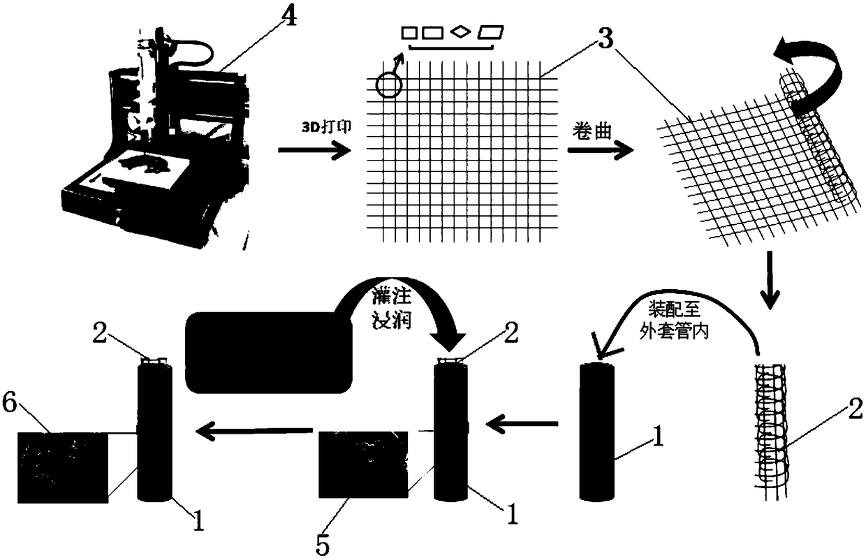
[0040] Such as figure 1 As shown, an embodiment of the peripheral nerve stent based on 3D printing of the present invention includes an outer sleeve 1 and a filler disposed in the outer sleeve 1, the filler is a three-dimensional skeleton 2 formed by crimping a gauze 3, and the gauze 3 is made of bio-ink, the outer sleeve 1 is made of electrospinning, the electrospinning is made of PLGA, and the gauze 3 is made of 3D printing.

[0041]Wherein, the bio-ink is a hydrogel prepared by modifying PEG with double bond end groups of acrylic acid; the three-dimensional skeleton is perfused or infiltrated with a solution that can promote axon growth, which contains β-NGF.

[0042] The preparation method of above-mentioned peripheral nerve stent, comprises the steps:

[0043] (1) Provide an outer sleeve made of electrospun material;

[0044] (2) Two-dimensional gauze was prepared by using an ink with dual sensitivity to temperature and ultraviolet light (bio-ink) and a 3D printing meth...

Embodiment 2

[0049] An embodiment of the peripheral nerve stent based on 3D printing of the present invention includes an outer sleeve 1 and a filler disposed in the outer sleeve 1. The filler is a three-dimensional skeleton 2 formed by crimping a gauze 3, and the gauze 3 adopts biological It is made of ink, the outer sleeve 1 is made of electrospinning, the electrospinning is made of PLLA and PCL, and the gauze is made of 3D printing method.

[0050] Among them, the bio-ink is hyaluronic acid modified by acrylic acid or norbornene; the three-dimensional framework is perfused or infiltrated with a hydrogel obtained by decellularizing and pulverizing peripheral nerves from pigs and digesting them with pepsin.

[0051] The preparation method of above-mentioned peripheral nerve stent, comprises the steps:

[0052] (1) Provide an outer sleeve made of electrospun material;

[0053] (2) Two-dimensional gauze was prepared by using ink with dual sensitivity to temperature and ultraviolet light (b...

Embodiment 3

[0058] An embodiment of the peripheral nerve stent based on 3D printing of the present invention includes an outer sleeve 1 and a filler disposed in the outer sleeve 1, the filler is a three-dimensional skeleton 2 formed by crimping gauze, and the gauze 3 uses bio-ink The outer casing 1 is made by electrospinning, the electrospinning is made by PTMC, and the gauze is made by 3D printing.

[0059] Among them, the bio-ink is tissue decellularized hydrogel; the tissue is pig-derived peripheral nerve, spinal cord, small intestinal submucosa, umbilical cord, placenta or amniotic membrane; the three-dimensional skeleton is perfused or infiltrated with β-NGF containing β-NGF that can promote axon growth. solution.

[0060] The preparation method of above-mentioned peripheral nerve stent, comprises the steps:

[0061] (1) Provide an outer sleeve made of electrospun material;

[0062] (2) Two-dimensional gauze was prepared by using an ink with dual sensitivity to temperature and ultr...


PUM


Abstract

The invention discloses a peripheral nerve stent based on 3D (three-dimensional) printing. The peripheral nerve stent comprises an outer cannula and a filler, wherein the filler is arranged in the outer cannula; the filler is a three-dimensional stent which is curved by a gauze net; the gauze net is made of biological ink. The invention also discloses a preparation method of the peripheral nerve stent. The peripheral nerve stent prepared by the preparation method has the advantages that the bionic elements of structure and composition of natural peripheral nerve are realized; on the basis of ensuring the biocompatibility, the outer cannula of electrospinning material can provide the full structure stability of the stent, the requirement on the ink forming strength can be reduced by the filler in the manufacturing process, the material selection range of the filler is expanded, and the ink can select more biological functions; by injecting or soaking special functional components into the stent, the corresponding biology characteristics are provided for the filler, such as acceleration of axone growth, and acceleration of vascularization.

Description

technical field [0001] The invention relates to the technical field of tissue engineering and bionic structures, in particular to a 3D printing-based peripheral nerve support and a preparation method thereof. Background technique [0002] The anatomical structure of the peripheral nerve is complex, which is divided into epineurium area, nerve fascicular area and interfascicular area, etc. There are also functional structures such as microvessels and connective tissue distributed in each area. In addition, the composition and spatial distribution of different regions are also very complex. Nerve repair materials (or catheters) currently reported in the literature are mainly prepared from a single homogeneous material (such as synthetic materials, natural materials, and acellular matrix materials). According to the principle of nerve regeneration, the structure and composition of the catheter have also been bionically designed (such as Gradient distribution of nerve growth fa...

Claims


Application Information

Patent Timeline
Patent Type & Authority Applications(China)
IPC IPC(8): A61L27/18A61L27/20A61L27/22A61L27/24A61L27/36A61L27/56A61L27/54B33Y10/00B33Y80/00
CPCA61L27/18A61L27/20A61L27/222A61L27/24A61L27/3633A61L27/3675A61L27/54A61L27/56A61L2300/414A61L2430/32B33Y10/00B33Y80/00C08L71/02C08L5/08
Inventor 王涛朱庆棠全大萍刘小林吴泽佳闫立伟
Owner THE FIRST AFFILIATED HOSPITAL OF SUN YAT SEN UNIV
Why Eureka
Try Eureka