Application of humanized brain-like organ to brain injury dyskinesia disease
A movement disorder and brain injury technology, applied in the field of brain science, to achieve the effect of good balance beam exercise ability and improvement of neuromuscular function
- Summary
- Abstract
- Description
- Claims
- Application Information
AI Technical Summary
Problems solved by technology
Method used
Image
Examples
Embodiment 1
[0035] Example 1: Culture of Brain Organoids
[0036] According to references (Nature 2013; 501:373-379 and Nat Protoc 2014; 9:2329-2340), after a series of explorations and improvements, the ultra-low adsorption well plate and rotary biogenerator culture system were adopted successively, and human pluripotent stem cells After successively undergoing germ layer differentiation, neuroectoderm differentiation, and neuroepithelial tissue formation, humanized brain organoids with 3D structures have been induced in rotary flasks. The culture stages are as follows.
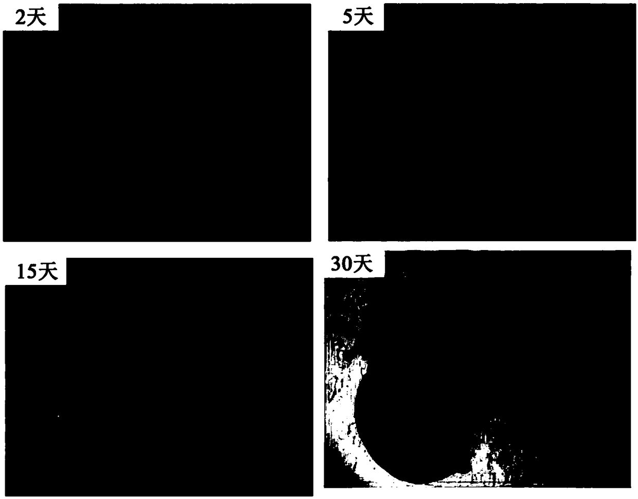
[0037] The first stage: In the ultra-low adsorption 96-well plate, according to the living cell density 9×10 4 cells / mL, inoculated 150 μL per well to obtain embryonic small body-like cell clusters ( figure 1 , 2 days shown).
[0038] The second stage: continue to induce differentiation, the embryonic body begins to show the characteristics of germ layer differentiation, and induces the initial neuroepithelial tissue ...
Embodiment 2
[0041] Example 2: Brain organoids used in brain injury transplantation
[0042] experimental method
[0043] 1. Construction of rat brain trauma model and brain transplantation
[0044] Adult SD rats, weighing about 300 g, were purchased from Shanghai Slack Experimental Animal Co., Ltd. The day before the operation, cyclosporine A (LC Labs C6000) 10 mg / kg was administered intraperitoneally. Rats were anesthetized by intraperitoneal injection, and then the head hair was shaved with a shaver, and fixed on a mouse brain stereotaxic apparatus. Wipe the rat scalp with 75% alcohol for disinfection, make a longitudinal incision about 4 cm long along the midline of the brain with a scalpel, separate the surface fascia of the skull, and fully expose the skull. In the right hemibrain of the rat, a rectangular skull window with a length of 1.5 cm and a width of 0.6 cm was drilled with a skull drill to expose the cortex of the rat. The mouse brain stereotaxic instrument was used as th...
PUM
 Abstract
Description
Claims
Application Information
 - R&D
- Intellectual Property
- Life Sciences
- Materials
- Tech Scout
- Unparalleled Data Quality
- Higher Quality Content
- 60% Fewer Hallucinations
Browse by: Latest US Patents, China's latest patents, Technical Efficacy Thesaurus, Application Domain, Technology Topic, Popular Technical Reports.
© 2025 PatSnap. All rights reserved.Legal|Privacy policy|Modern Slavery Act Transparency Statement|Sitemap|About US| Contact US: help@patsnap.com


