Functional collagen biological material capable of inducing tympan regeneration and preparation method thereof

A collagen biology, collagen membrane technology, applied in tissue regeneration, chemical instruments and methods, antibody mimics/scaffolds, etc., can solve the problems of expanding incision, increasing trauma, increasing the difficulty of material acquisition, etc., to achieve the effect of promoting repair

Inactive Publication Date: 2018-12-25
INST OF GENETICS & DEVELOPMENTAL BIOLOGY CHINESE ACAD OF SCI
View PDF6 Cites 0 Cited by
  • Summary
  • Abstract
  • Description
  • Claims
  • Application Information

AI Technical Summary

Problems solved by technology

Among them, the autologous graft material is the most commonly used, with a high success rate, but because of the need to enlarge the inci

Method used


Image

  • Functional collagen biological material capable of inducing tympan regeneration and preparation method thereof
  • Functional collagen biological material capable of inducing tympan regeneration and preparation method thereof

Examples


Embodiment 1

[0059] Embodiment 1, the preparation of collagen film (CM)

[0060] 1. Take the dermis tissue of isolated bovine skin (thickness is 1 mm), soak and wash in deionized water 10 times, 10 minutes each time.

[0061] 2. Treat the dermal tissue treated in step 1 in turn according to the following steps:

[0062] (1) Place in 1% by mass sodium lauryl sulfate aqueous solution, let stand at 16° C. for 6 hours, then soak and wash in deionized water for 5 minutes.

[0063] (2) Place in 1% by mass Tween-80 aqueous solution, let stand at 16° C. for 24 hours, then soak and wash in deionized water for 5 minutes.

[0064] (3) Place in 2M aqueous potassium hydroxide solution and let stand at 16°C for 20 minutes.

[0065] (4) Soak and wash in deionized water for one hour (change the liquid 20 times).

[0066] Freeze-dried to obtain a collagen film.

Embodiment 2

[0067] Embodiment 2, preparation of basic fibroblast growth factor

[0068] 1. Construction of recombinant expression vector of basic fibroblast growth factor with collagen binding ability

[0069] 1. Artificially synthesize the double-stranded DNA molecule shown in the 58th-459th nucleotide of the 5' end of the sequence 1 to obtain the CBD-mFGF DNA fragment.

[0070] 2. Digest the CBD-mFGF DNA fragment obtained in step 1 with restriction endonucleases NdeI and XhoI.

[0071] 3. Digest the pET-28a(+) vector with restriction endonucleases NdeI and XhoI, and recover the vector backbone of about 5284bp.

[0072] 4. Ligate the DNA fragment obtained in step 2 with the vector backbone obtained in step 3 to obtain the recombinant vector pET28a-CBD-mFGF. According to the sequencing results, the structure of the recombinant vector pET28a-CBD-mFGF is described as follows: the small fragment between the NdeI and XhoI restriction sites of the pET-28a(+) vector is replaced by the 5' end ...

Embodiment 3

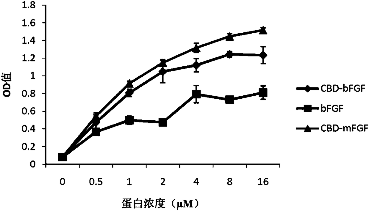
[0092] Embodiment 3, detection of binding ability of basic fibroblast growth factor and collagen membrane (CM)

[0093] The protein solutions to be tested are: the CBD-mFGF protein solution, the CBD-bFGF protein solution and the bFGF protein solution prepared in Example 2.

[0094] 1. Get the collagen film prepared in Example 1 (collagen film area 0.32cm 2 , thickness 1 mm), immersed in 200 μL of blocking solution, and blocked for 2 hours at 37° C. and 100 rpm. Blocking solution: PBS buffer (pH7.4) containing 2.5% (volume percent) BSA and 0.1% (volume percent) Tween20. After blocking, the collagen membrane was soaked in PBS buffer (pH7.4) and washed 4 times.

[0095] 2. Dilute each protein solution to be tested into protein dilutions with different concentrations (0 μM, 0.5 μM, 1 μM, 2 μM, 4 μM, 8 μM and 16 μM) with PBS buffer (pH 7.4). Each protein dilution was dropped onto the collagen membrane treated in step 1 (20 μL per membrane), and incubated at 37°C for 30 minutes. ...


PUM

PropertyMeasurementUnit
Thicknessaaaaaaaaaa

Abstract

The invention discloses a functional collagen biological material capable of inducing tympan regeneration and a preparation method thereof. The invention provides protein which comprises a CBD sectionand an mFGF section, an amino acid sequence of the CBD section is 22th-28th of N end of a sequence 2 in a sequence table, and an amino acid sequence of the mFGF section is 36th-149th of the N end ofthe sequence 2 in the sequence table. The invention further provides a preparation method of the functional collagen biological material. The preparation method includes following steps: loading protein onto a collagen film to obtain the collagen biological material. The collagen film which can be specifically combined with basic fibroblast growth factor is prepared, microenvironment for promotingtympan regeneration is built, and recovery after tympan perforation is promoted.

Description

technical field [0001] The invention relates to a functional collagen biomaterial for inducing tympanic membrane regeneration and a preparation method thereof. Background technique [0002] Perforation of the tympanic membrane caused by infection and trauma is a common clinical disease. Usually, the spontaneous recovery rate of tympanic membrane perforation is about 78.7%, and the average treatment period is about 1-3 months. The perforated tympanic membrane heals in the medial tympanic mucosal epithelium through migration of epithelial cells on the tympanic surface, but the central fibrous layer is often absent. If the blood supply is poor, the surface epithelial hyperplasia migration is weakened or the perforation is too large, the fibrous tissue is insufficient to provide support for epithelial migration, and the tympanic membrane will not heal itself. Tympanic membrane repair is a common method. At present, the materials used clinically for tympanic membrane repair can...

Claims


Application Information

Patent Timeline
IPC IPC(8): C07K19/00C12N15/62A61L27/24
CPCA61L27/24A61L2430/14C07K14/503C07K2319/00
Inventor 戴建武赵燕南侯祥林陈冰施勤
Owner INST OF GENETICS & DEVELOPMENTAL BIOLOGY CHINESE ACAD OF SCI
Why Eureka
Try Eureka