Method for building three-dimensional analog brain development models on basis of micro-fluidic chips

A technology of microfluidic chips and construction methods, applied in biochemical equipment and methods, specific-purpose bioreactors/fermenters, tissue cell/virus culture devices, etc.

Active Publication Date: 2018-12-25
DALIAN INST OF CHEM PHYSICS CHINESE ACAD OF SCI
View PDF5 Cites 5 Cited by
  • Summary
  • Abstract
  • Description
  • Claims
  • Application Information

AI Technical Summary

Problems solved by technology

However, the combination of microfluidic technology and brain-like technology t

Method used


Image

  • Method for building three-dimensional analog brain development models on basis of micro-fluidic chips
  • Method for building three-dimensional analog brain development models on basis of micro-fluidic chips
  • Method for building three-dimensional analog brain development models on basis of micro-fluidic chips

Examples


Embodiment 1

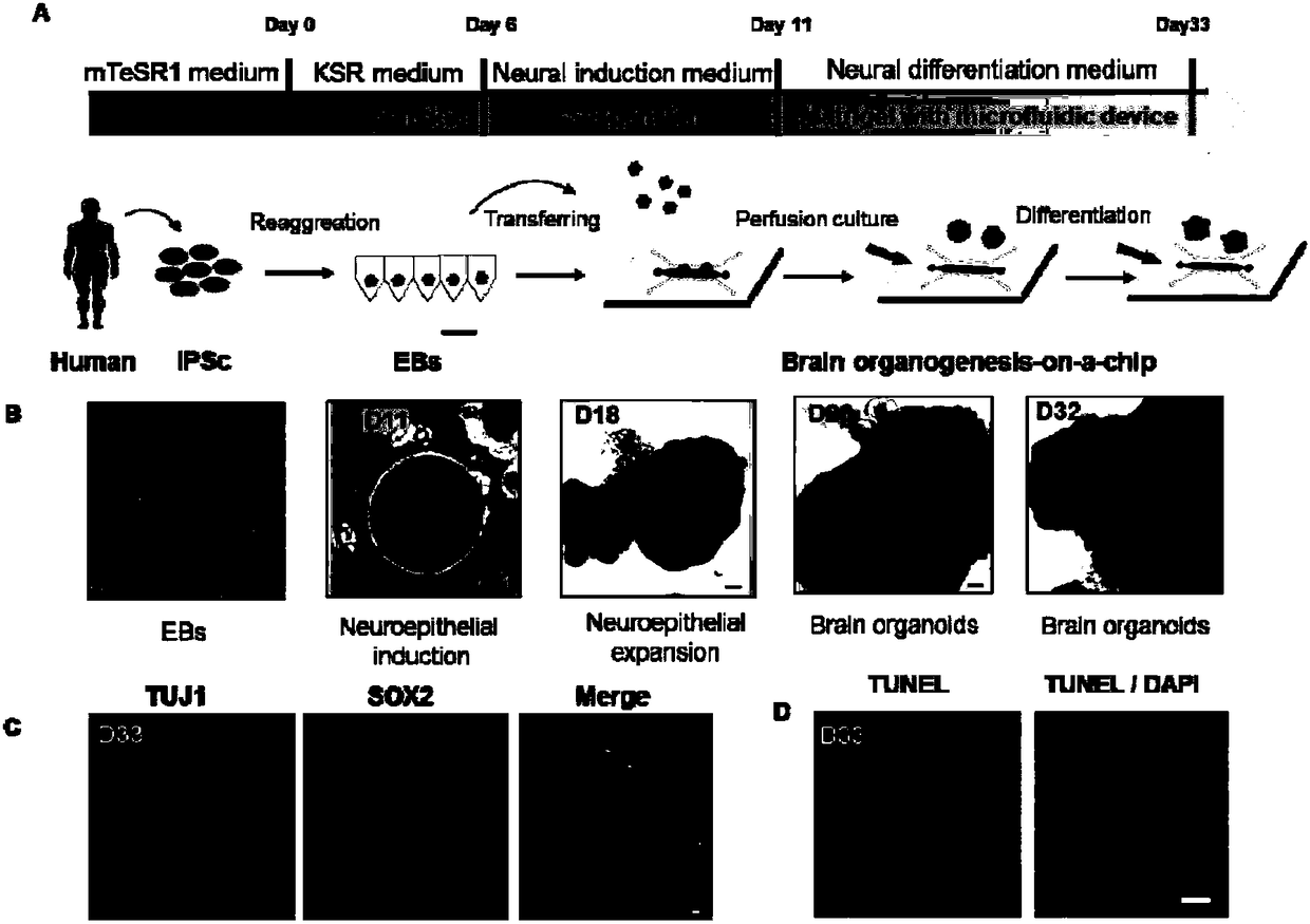
[0042] Development of HiPSC-derived brain-like cells on a microfluidic chip

[0043] The steps are mainly divided into three parts: the preparation of the microfluidic chip, the formation of embryoid body EBs and the development of the brain in the microfluidic chip.

[0044] Step (1) The preparation of the microfluidic chip, specifically: the microfluidic chip is formed by irreversible sealing of the upper and lower layers, and the materials of the upper and lower layers are polymerized polydimethylsiloxane, a transparent and breathable biocompatible material. things. Then the upper and lower layers of polymer materials are treated with oxygen plasma for 60 seconds for irreversible sealing. After sealing, it is sterilized by high temperature and high pressure for standby use.

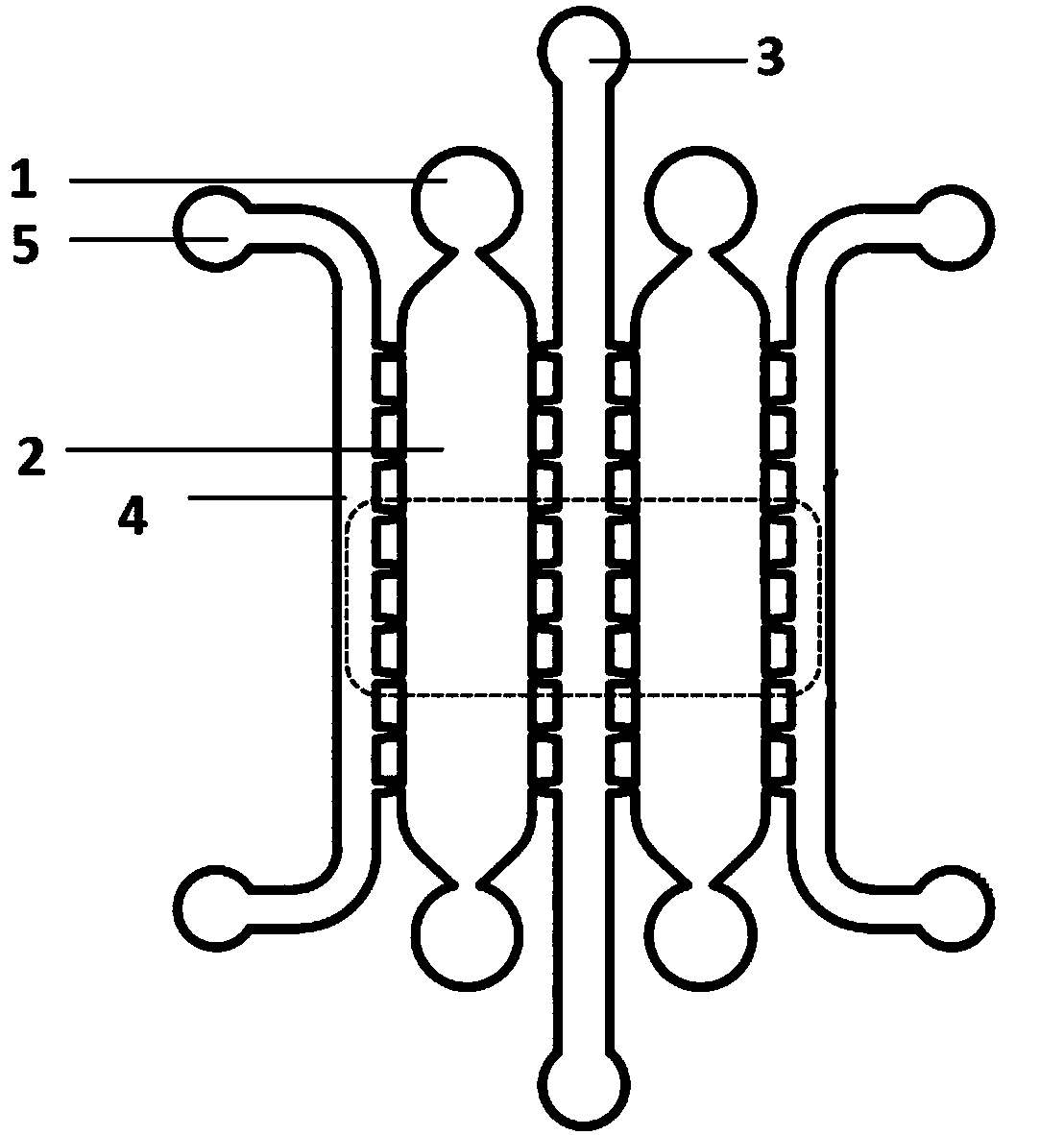
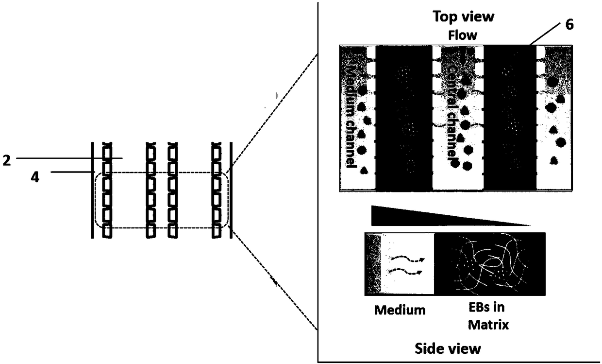
[0045] Microfluidic chips such as figure 1 As shown, it includes an extracellular matrix suspension inlet (1), a three-dimensional channel (2), a two-dimensional perfusion channel (3), a two-dimensi...

Embodiment 2

[0050] Expression of associated proteins in different brain regions during development.

[0051] The static and perfusion cultured brains grown on the microfluidic chip for 33 days were taken for cryosection respectively. The method was as follows: 4% paraformaldehyde was used to fix the cells for 20 minutes, washed with PBS buffer three times, each time for 10 minutes; 30% sucrose Dehydrate overnight at 4°C; embedding in OCT, store at room temperature for 30 minutes, and solidify at 80°C; freeze sections with a thickness of 10-20 μm, and attach them to electrostatically adsorbed glass slides. Immunofluorescence staining was then carried out. The method was as follows: soak the slides with sections in PBS buffer for 5 minutes; act with 0.1% triton X-100 porogen for 10 minutes, rinse once with PBS buffer for 5 minutes; goat blocking serum React at room temperature for 1 hour; primary antibodies (PAX6, PAX2, NESTIN, TUJ1, SOX2) were diluted 1:400, incubated overnight at 4°C, and...

Embodiment 3

[0053] Expression of neuron-associated proteins in different cortical layers during development.

[0054] The static and perfusion cultured brains grown on the microfluidic chip for 33 days were taken for cryosection respectively. The method was as follows: 4% paraformaldehyde was used to fix the cells for 20 minutes, washed with PBS buffer three times, each time for 10 minutes; 30% sucrose Dehydrate overnight at 4°C; embedding in OCT, store at room temperature for 30 minutes, and solidify at 80°C; freeze sections with a thickness of 10-20 μm, and attach them to electrostatically adsorbed glass slides. Immunofluorescence staining was then carried out. The method was as follows: soak the slides with sections in PBS buffer for 5 minutes; act with 0.1% triton X-100 porogen for 10 minutes, rinse once with PBS buffer for 5 minutes; goat blocking serum Effect at room temperature for 1 hour; primary antibody (TBR1, CTIP2, BRN2) was diluted 1:400, incubated overnight at 4°C, washed 3 ...


PUM

PropertyMeasurementUnit
Diameteraaaaaaaaaa
Depthaaaaaaaaaa

Abstract

The invention discloses a method for building three-dimensional analog brain development models on the basis of micro-fluidic chips. The method includes main steps of preparing the micro-fluidic chips; forming embryoid bodies (EBs); differentiating and developing analog brains in the micro-fluidic chips. The method has the advantages that the novel engineered three-dimensional analog brain development models can be built by the aid of the method and are micro-reactors with perfusion functions on the basis of micro-fluidic chip technologies; in-vivo brain development basic principles and engineering technologies are combined with one another, and brain early development micro-environments can be effectively simulated; the method is low in cost and reagent dosage and easy to operate, in-situtracking and real-time monitoring can be carried out, animal models and the traditional two-dimensional culture modes can be replaced, brain early development can be simulated to a certain extent, apowerful novel platform can be provided to the aspects such as in-vitro brain development simulation, pathological research on abnormal neural development, medicine screening and toxicity detection, and the like.

Description

technical field [0001] The invention relates to the fields of microfluidic chip technology and tissue engineering, in particular to a method for constructing a brain-inspired development model based on a microfluidic chip. Background technique [0002] Brain-like development from various stem cell sources has been effectively developed in recent years, including neural stem cells, intestinal stem cells, embryonic stem cells, and induced pluripotent stem cells (iPSCs). The formation of brain-like cells originates from embryoid body EBs, which are specifically differentiated from stem cells and self-assembled into multicellular aggregates with a three-dimensional structure, which are further developed into tissues with certain structural and functional specificities in vitro, which simulates to a certain extent Corresponding brain development process. Studies have shown that iPSCs can differentiate into specific cell types and have strong self-assembly ability to form a varie...

Claims


Application Information

Patent Timeline
IPC IPC(8): C12N5/073C12M3/00
CPCC12M21/08C12M23/16C12N5/0603
Inventor 秦建华王亚清王丽
Owner DALIAN INST OF CHEM PHYSICS CHINESE ACAD OF SCI
Why Eureka
Try Eureka