Preparation method of biovalve

A biological valve and biological tissue technology, applied in the field of medical devices, can solve problems affecting the safety of artificial biological valves, and achieve the effects of improving anti-calcification performance, reducing voids, and reducing deposition

Inactive Publication Date: 2019-01-04
SHANGHAI MICROPORT CARDIOFLOW MEDTECH CO LTD
View PDF7 Cites 9 Cited by
  • Summary
  • Abstract
  • Description
  • Claims
  • Application Information

AI Technical Summary

Problems solved by technology

[0007] In summary, it can be seen that there is currently no method for preparing a bioprosthetic valve, which can avoid the loss of GAG

Method used


Image

  • Preparation method of biovalve
  • Preparation method of biovalve
  • Preparation method of biovalve

Examples


preparation example Construction

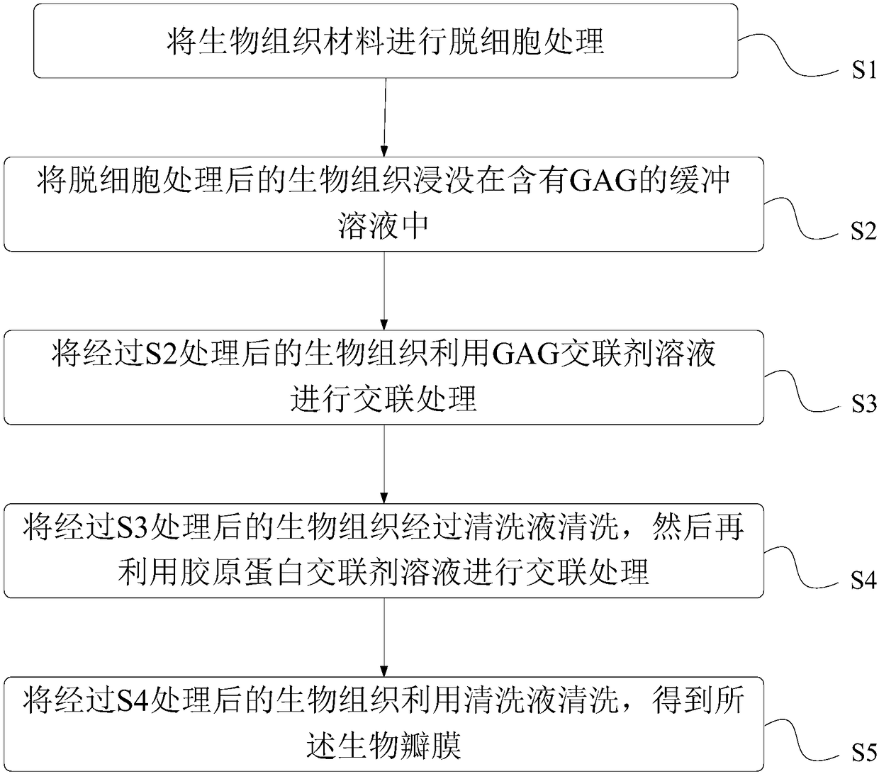
[0052] figure 1 It is a schematic flowchart of a method for preparing a biological valve provided by the present invention.

[0053] The preparation method provided by the invention comprises the following steps:

[0054] S1: Decellularize the biological tissue material to obtain the decellularized biological tissue.

[0055] Wherein, the biological tissue material can be the same or heterogeneous biological tissue material, for example, any one selected from bovine pericardium, porcine pericardium, equine pericardium and porcine aortic valve. The decellularization method for treating biological tissue is not particularly limited, as long as it can remove cells in the tissue and related degradation enzymes, phospholipids and cell membrane components in the tissue that cause calcification, and improve the anti-calcification performance of the tissue. The methods of decellularization treatment include: hypotonic shaking treatment method, surfactant method, repeated freezing an...

Embodiment 1

[0084] A kind of preparation method of artificial biological valve comprises the steps:

[0085] S1: Bovine pericardium tissue obtained from the slaughterhouse, after fat stripping, trimming and washing, was shaken with hypotonic solution (pH 7.4) and 1% (w / v) octylglucopyranoside (Sigma-Aldrich Co .LLC.) combined with decellularization.

[0086] S2: Submerge the treated bovine pericardium in HEPES (Sigma-Aldrich Co.LLC.) solution (pH 7.4) containing 0.02% (w / v) dermatan sulfate (Sigma-Aldrich Co.LLC.) for 24h, Remove the tissue.

[0087] S3: The tissue treated in S2 above was treated with 0.006% (w / v) N-hydroxy-succinimide (Sigma-Aldrich Co. LLC.) and 0.03% (w / v) 1-cyclohexyl-2-mol Morpholineethanesulfonic acid (Shanghai Crystal Pure Biochemical Technology Co., Ltd.) solution (pH value 5.5) of morpholine ethyl carbodiimide p-toluenesulfonate (Shanghai Crystal Pure Biochemical Technology Co., Ltd.) was cross-linked for 24 hours.

[0088] S4: The above-mentioned tissues afte...

Embodiment 2

[0099] The preparation method of the artificial biological valve in this embodiment is similar to that of Embodiment 1, specifically including the following steps:

[0100] S1: Porcine pericardium tissue was obtained from a slaughterhouse. After fat stripping, trimming and cleaning, 1% TritonX-100 (w / v) (Shanghai Jingchun Biochemical Technology Co., Ltd.) was used for decellularization.

[0101] S2: Submerge the treated porcine pericardium in HEPES solution (pH 7.4) containing 0.008% (w / v) dermatan sulfate and 0.002% chondroitin sulfate (Sigma-Aldrich Co.LLC.) for 12 hours, and remove the tissue .

[0102] S3: The tissue treated in S2 above was treated with 0.02% (w / v) 4-(4,6-dimethoxytriazin-2-yl)-4-methylmorpholine hydrochloride (Suzhou Haofan Biological Co., Ltd.) morpholine propanesulfonic acid (Shanghai Jingchun Biochemical Technology Co., Ltd.) solution (pH value 5.0) cross-linked for 24h.

[0103] S4: Wash the above-mentioned tissues cross-linked by S3 with phosphate ...


PUM

PropertyMeasurementUnit
Molecular weightaaaaaaaaaa

Abstract

The invention provides a preparation method of a biovalve. The method comprises the steps that S1, decellularization is conducted on a biological tissue material to obtain biological tissues on whichdecellularization is conducted; S2, the biological tissues on which decellularization is conducted are immersed in a buffering solution containing GAG; S3, cross-linking treatment is conducted on thebiological tissues treated in S2 by using GAG cross-linking agent solution; S4, the biological tissues treated in S3 are washed by cleaning liquid, and cross-linking treatment is conducted by using collagen protein cross-linking agent; S5, the biological tissues treated in S4 are cleaned by using the cleaning liquid to obtain the biovalve. By means of the biovalve obtained through the treated biological tissues, tissue wrinkling can be significantly relieved or eliminated to reduce stress generated by tissue bending to increase the smoothness of the tissues, improve the using rate of tissue materials and greatly reduce production cost.

Description

technical field [0001] The invention belongs to the field of medical instruments and relates to a preparation method of a biological valve. Background technique [0002] Artificial biological valves are mainly prepared from animal-derived biological tissue materials such as porcine aortic valves, bovine pericardium, and porcine pericardium. These biological tissue materials are mainly composed of collagen fibers, elastic fibers, and GAG. [0003] Animal-derived biological tissue materials are generally fixed with glutaraldehyde in the process of preparing artificial biological valves, but the tissue after glutaraldehyde fixation will harden, and it is easy to form wrinkles when the tissue is bent. After the prepared bioprosthetic valve is implanted in the body, the valve leaflet will bend sharply along the valve frame to form folds during the opening and closing process, which will cause the collagen fibers in the valve leaflet to be easily damaged. Once the collagen fibers...

Claims


Application Information

Patent Timeline
IPC IPC(8): A61L27/36A61L27/50
CPCA61L27/3625A61L27/3687A61L27/50A61L2430/20A61L2430/40
Inventor 董教明桂宝珠张泉娟陈国明李雨
Owner SHANGHAI MICROPORT CARDIOFLOW MEDTECH CO LTD
Why Eureka
Try Eureka