Dual fluorescent microsphere immunological detection method for pseudorabies virus gE and gB IgG antibodies

A technology of porcine pseudorabies virus and fluorescent microspheres, which is applied in measuring devices, scientific instruments, biological testing, etc., can solve the problems of single method and high cost, and achieve the effects of good repeatability, high sensitivity and great application value

Active Publication Date: 2019-02-05
SOUTH CHINA AGRI UNIV
View PDF9 Cites 4 Cited by
  • Summary
  • Abstract
  • Description
  • Claims
  • Application Information

AI Technical Summary

Problems solved by technology

However, at present, the single ELISA method of gE and gB protein IgG antibodies is mainly used clinically to detect and analyze the infection of the virus and the level of protective antibodies, which has the disadvantages of high cost and single method

Method used


Image

  • Dual fluorescent microsphere immunological detection method for pseudorabies virus gE and gB IgG antibodies
  • Dual fluorescent microsphere immunological detection method for pseudorabies virus gE and gB IgG antibodies
  • Dual fluorescent microsphere immunological detection method for pseudorabies virus gE and gB IgG antibodies

Examples


Embodiment 1

[0072] Embodiment 1 The amplification of PRV gE and gB gene

[0073] 1. Experimental operation

[0074] The upstream and downstream primers were designed respectively according to the whole genome sequence of herpes virus KU056477.1 and the whole genome sequence of herpes type 1 Guangdong isolate with ID KT948041.1 provided by NCBI GenBank (see Table 1). Since it is impossible to design suitable primers for cloning the full-length gene directly using DNA as a template, two sets of primers were designed for two rounds of cloning to obtain PRV gE and gB truncated genes.

[0075] Table 1 Primers:

[0076]

[0077]

[0078] In the first round of amplification, gEF1, gER1, gBF1, and gBR1 were used to amplify PRV gE and gB genes, respectively.

[0079] In the first round of amplification, gEF2, gER2, gBF2, and gBR2 were used to amplify PRV gE and gB genes, respectively. The second round of primers contains EcoR I and HindШ restriction sites, and the bold content in the tabl...

Embodiment 2

[0083] Construction and identification of embodiment 2 recombinant plasmids pMAL-c5x PRV gE, pMAL-c5x PRV gB

[0084] 1. Experimental operation

[0085] The obtained PRV gE and PRV gB gene fragments were respectively ligated with the pMAL-c5X expression vector after double digestion with EcoR I and HindШ restriction endonucleases, and named as pMAL-c5x PRV gE and pMAL-c5x PRV gB respectively. After the ligation product was transformed, the single colony picked was subjected to the identification of the recombinant plasmid after expansion: (1) PCR; (2) double digestion of the two recombinant plasmids with restriction endonucleases EcoR I and HindШ; ( 3) Send the extracted plasmid to Sangon Biotechnology Co., Ltd. for sequencing.

[0086] 2. Experimental results

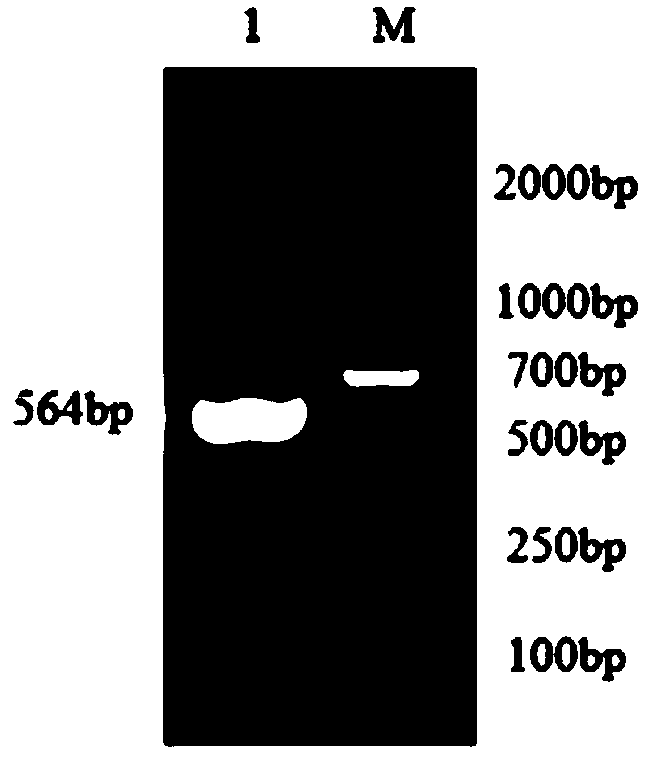
[0087] After PCR, gel electrophoresis obtained specific bands of about 546bp and 609bp, respectively, which were consistent with the expected size.

[0088] After double enzyme digestion, gel electrophoresis, pMAL-c...

Embodiment 3

[0090] Example 3 Detection of antigen induced expression and soluble analysis, Western-blot verification

[0091] 1. Experimental operation

[0092] (1) Induced expression of antigen

[0093] The positive recombinant plasmids successfully sequenced were transformed into E. coli BL21(DE3) Escherichia coli, and the inducer isopropylthiogalactopyranoside (IPTG) was added at a final concentration of 0.3mM, and the expression was induced in a shaker at 16°C for 8h.

[0094] (2) Solubility analysis of antigen

[0095] Centrifuge the induced bacterial solution at 8000rpm / min for 10min, discard the supernatant, and resuspend the bacterial cells with PBS. The resuspended cells were ultrasonically disrupted in an ice bath, 200W, working for 3 sec, intermittent for 5 sec, until the bacterial liquid became clear. The bacterial solution was centrifuged at 12000rpm at 4°C for 20min, the supernatant and the precipitate were collected respectively, and the precipitate was resuspended in PBS ...


PUM

PropertyMeasurementUnit
Sensitivityaaaaaaaaaa

Abstract

The invention discloses a dual fluorescent microsphere immunological detection method for pseudorabies virus gE and gB IgG antibodies. The detection method is based on a liquid protein chip technology-based carboxylated fluorescent microsphere group for detecting pseudorabies virus antibodies; the group comprises carboxylated fluorescent microspheres coupled with gE truncated proteins and gB truncated proteins, respectively; and the amino acid sequences of gE and gB truncated proteins are shown in SEQ ID NOs: 2 and 4, respectively. The dual fluorescent microsphere immunological detection method for simultaneously detecting PRV gE and gB IgG antibodies is good in reproducibility, high in sensitivity and good in specificity, and has no cross-reaction with other common porcine virus-positiveserums. The method can be used for the rapid differential diagnosis of pseudorabies virus wild-type infected pigs and vaccine-vaccinated pigs and the detection of protective antibodies, thereby providing an important method for the monitoring of swine virus diseases, having a great application value and being worthy of large-scale promotion.

Description

technical field [0001] The invention relates to the technical field of virus detection, more specifically, to a double fluorescent microsphere immunological detection method of pseudorabies virus gE and gB IgG antibodies. Background technique [0002] Pseudorabies virus (PRV) is a member of the family Herpesviridae and the subfamily Alphaherpesvirinae. Pigs are the natural host and source of infection of PRV, which can cause fatal encephalitis in newborn pigs, respiratory disorder in fattening pigs, reproductive failure in breeding pigs, abortion, stillbirth, and mummification in pregnant sows. Adult pigs have no obvious symptoms after infection, showing recessive infection. After tolerance, they will cause long-term poisoning and detoxification, becoming the most dangerous source of infection and bringing huge economic losses to the pig industry. [0003] Since the PRV gE gene is expressed in all wild strains; at the same time, the gE deletion vaccine is safer than other g...

Claims


Application Information

Patent Timeline
Patent Type & Authority Applications(China)
IPC IPC(8): G01N33/68G01N33/569G01N33/543G01N33/533
CPCG01N33/533G01N33/54313G01N33/56994G01N33/6857G01N2333/032
Inventor 王衡张桂红曾梦冀池海
Owner SOUTH CHINA AGRI UNIV
Why Eureka
Try Eureka