Preparation method and application of magnetic nanoparticles for capturing exosomes in blood
A technology of magnetic nanoparticles and micro-nanoparticles, which is applied in the field of tumor detection, can solve the problems of low processing capacity, long processing time, and low repetition rate, and achieve the effects of short processing time, small sample size, and easy operation
- Summary
- Abstract
- Description
- Claims
- Application Information
AI Technical Summary
Problems solved by technology
Method used
Image
Examples
Embodiment 1
[0020] A method for preparing magnetic nanoparticles for capturing exosomes in blood, comprising the following steps:
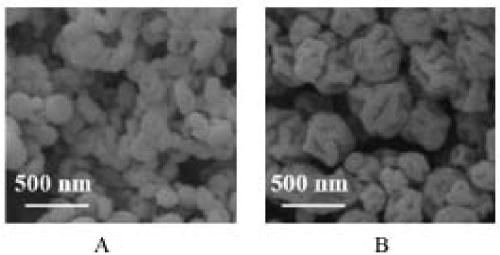
[0021] (1) 1.8mol ferric chloride hexahydrate (FeCl 3 ·6H 2 O) and 1 mol of ferrous chloride tetrahydrate (FeCl 2 4H 2 O) After mixing, dissolve in 100 mL of distilled water, stir and mix well, heat to 70°C, then slowly add NH with a concentration of 0.4mol / L 3 ·H 2 O solution, when the pH of the solution rises to 6-7, the iron salt hydrolyzes to produce a large amount of Fe 3 o 4 Crystal particles; continue to add NH dropwise 3 ·H 2 0 until the pH rises to 9-10, the mixed solution gradually turns black, and the hydrolysis tends to be complete; after the hydrolysis is basically completed, slowly add 2 mL of surfactant-cetyltrimethylammonium bromide (Cetyltrimethylammonium Bromide, CTAB), after stirring for 30 min, the product was collected by centrifugation; the product was repeatedly washed with distilled water until PH = 7.0, the supernatant was rem...
Embodiment 2
[0026] With above-mentioned embodiment 1, its difference is:
[0027] In step (1): mix ferric chloride hexahydrate and ferrous chloride tetrahydrate at a molar ratio of 1.6:1, dissolve in distilled water to obtain a mixed solution, stir and mix well, heat to 55°C, and then add dropwise at a concentration of 0.3mol / L of NH 3 ·H 2 O solution, when the hydrolysis of the mixed solution is completed, slowly add 1.5 mL of surfactant to the mixed solution, centrifuge to collect the product after stirring, wash the product repeatedly until pH = 7.0, remove the supernatant, take the lower layer and dry it in vacuum at 50 °C More than 16h, get nano-Fe 3 o 4 particle;
[0028] In step (2): take the nano-Fe obtained in step (1) 3 o 4 The particles were dispersed in a solution mixed with ethanol and distilled water at a ratio of 3:1 by volume. After ultrasonic treatment, a solution of tetraethyl orthosilicate with a concentration of 0.15 mol / L was added, and 6 mL was gradually added...
Embodiment 3
[0030] With above-mentioned embodiment 1, its difference is:
[0031] In step (1): mix ferric chloride hexahydrate and ferrous chloride tetrahydrate at a molar ratio of 2.0:1, dissolve in distilled water to obtain a mixed solution, stir and mix well, heat to 85°C, and then add dropwise at a concentration of 0.5mol / L NH 3 ·H 2 O solution, when the hydrolysis of the mixed solution is completed, slowly add 2.5 mL of surfactant to the mixed solution, centrifuge to collect the product after stirring, wash the product repeatedly until pH = 7.0, remove the supernatant, take the lower layer and dry it in vacuum at 70 °C More than 16h, get nano-Fe 3 o 4 particle;
[0032] In step (2): take the nano-Fe obtained in step (1) 3 o 4 The particles were dispersed in a solution mixed with ethanol and distilled water at a ratio of 5:1 by volume. After ultrasonic treatment, a solution of tetraethyl orthosilicate with a concentration of 0.25 mol / L was added, and 7.5 mL was gradually added ...
PUM
| Property | Measurement | Unit |
|---|---|---|
| diameter | aaaaa | aaaaa |
| particle diameter | aaaaa | aaaaa |
| particle diameter | aaaaa | aaaaa |
Abstract
Description
Claims
Application Information
 - R&D
- Intellectual Property
- Life Sciences
- Materials
- Tech Scout
- Unparalleled Data Quality
- Higher Quality Content
- 60% Fewer Hallucinations
Browse by: Latest US Patents, China's latest patents, Technical Efficacy Thesaurus, Application Domain, Technology Topic, Popular Technical Reports.
© 2025 PatSnap. All rights reserved.Legal|Privacy policy|Modern Slavery Act Transparency Statement|Sitemap|About US| Contact US: help@patsnap.com


