Nano-hydroxyapatite-coated porous titanium scaffold and its preparation method and application
A nano-hydroxyapatite and porous titanium technology, applied in the field of biomedical materials, can solve the problems of less tumor cell apoptosis and no local tumor recurrence, and achieve inhibition of tumor growth, good apoptosis of cancer cells, and good application prospects. Effect
- Summary
- Abstract
- Description
- Claims
- Application Information
AI Technical Summary
Problems solved by technology
Method used
Image

Examples
Embodiment 1
[0036] Example 1. Nano-hydroxyapatite-coated porous titanium stent
[0037] S1. Preparation of porous titanium substrate material
[0038] (1) Import the CT image into the simpleware 3D image software to obtain a 3D image of the target bone tissue. The average pore column is 500 μm and the average pore diameter is 300 μm. The image is filled and expanded with cubic porous units to obtain a personalized porous connected 3D digital model ;
[0039] (2) using a 3D printer, using titanium alloy as a raw material, and printing the porous titanium alloy stent according to the porous connected three-dimensional digital model designed in step (1);
[0040] S2, acid-base treatment is carried out to the porous titanium base material prepared in step S1, and a nano-scale microporous network TiO with a thickness of 0.5 μm is prepared on the pore wall and the surface of the skeleton 2 layer, the nanoscale microporous network TiO 2 layer porous titanium substrate material;
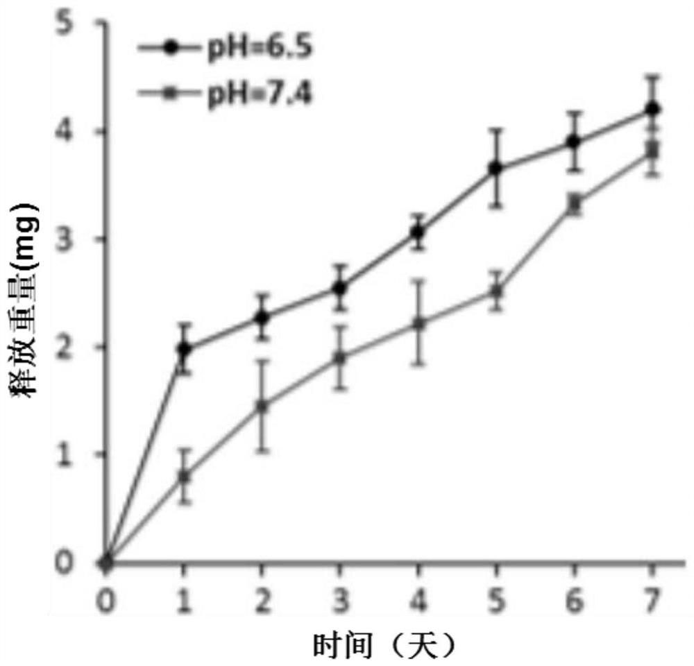
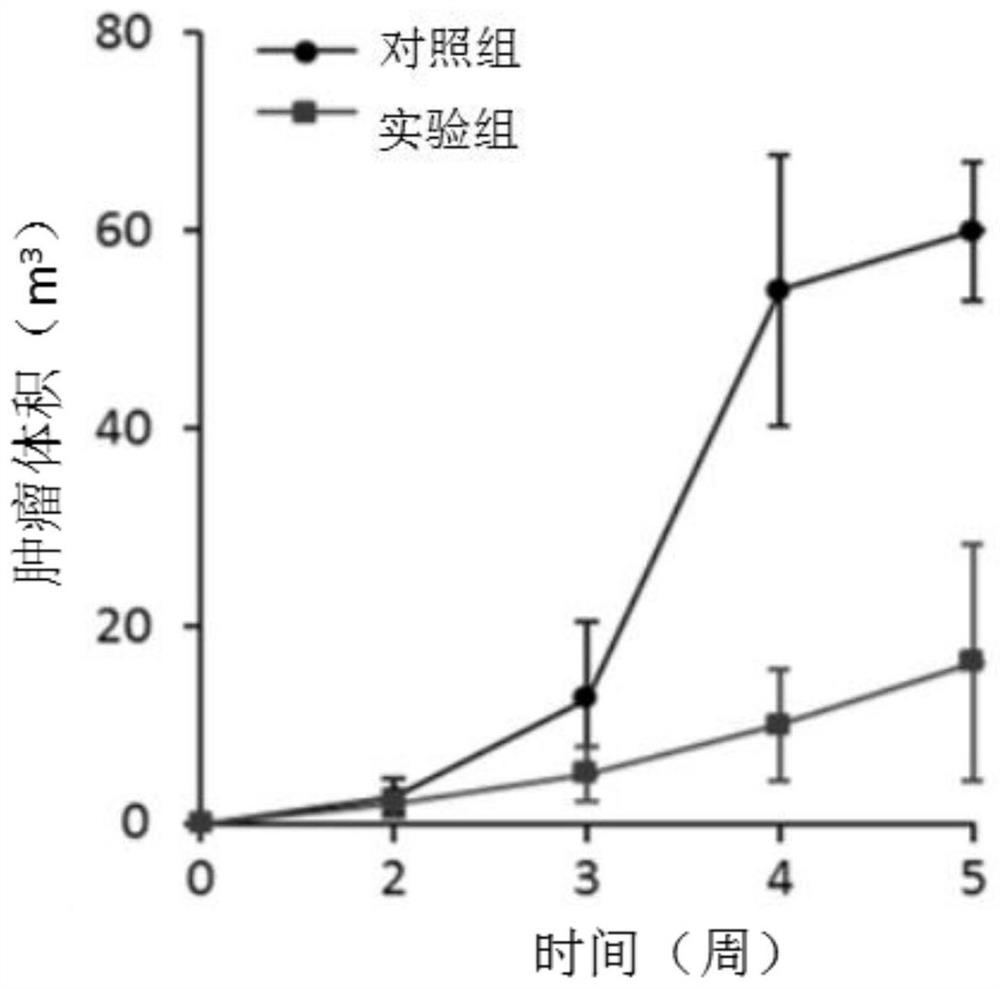
[0041] S3. Pr...
Embodiment 2
[0046] Example 2, nano-hydroxyapatite-coated porous titanium stent
[0047] S1. Preparation of porous titanium substrate material
[0048] (1) Import the CT image into the simpleware 3D image software to obtain a 3D image of the target bone tissue. The average pore column is 600 μm and the average pore diameter is 800 μm. The image is filled and expanded with octahedral porous units to obtain a personalized porous connected 3D digital image. Model;
[0049] (2) Use a 3D printer, use titanium alloy as raw material, and print a porous titanium alloy stent according to the design model;
[0050] S2. Alkaline heat treatment is performed on the porous titanium base material prepared in step S1, and a nanoscale microporous network TiO with a thickness of 2.0 μm is formed on the pore wall and the surface of the skeleton. 2 layer, the nanoscale microporous network TiO 2 layer porous titanium substrate material;
[0051] S3. Prepare nano-hydroxyapatite particle slurry by wet chemic...
Embodiment 3
[0056] Example 3, nano-hydroxyapatite-coated porous titanium stent
[0057] S1. Preparation of porous titanium substrate material
[0058] (1) Import the CT image into the simpleware 3D image software to obtain a 3D image of the target bone tissue. The average pore column is 400 μm and the average pore diameter is 600 μm. The image is filled and expanded with helical dodecahedral porous units to obtain personalized porous connectivity. 3D digital model;
[0059] (2) Use a 3D printer, use titanium alloy as raw material, and print a porous titanium alloy stent according to the design model;
[0060] S2, acid-base treatment is carried out to the porous titanium base material prepared in step S1, and a nanoscale microporous network TiO with a thickness of 1.2 μm is formed on the pore wall and the surface of the skeleton 2 layer, the nanoscale microporous network TiO 2 layer porous titanium substrate material;
[0061] S3. Prepare nano-hydroxyapatite particle slurry by wet chem...
PUM
| Property | Measurement | Unit |
|---|---|---|
| thickness | aaaaa | aaaaa |
| pore size | aaaaa | aaaaa |
| compressive strength | aaaaa | aaaaa |
Abstract
Description
Claims
Application Information
 - R&D
- Intellectual Property
- Life Sciences
- Materials
- Tech Scout
- Unparalleled Data Quality
- Higher Quality Content
- 60% Fewer Hallucinations
Browse by: Latest US Patents, China's latest patents, Technical Efficacy Thesaurus, Application Domain, Technology Topic, Popular Technical Reports.
© 2025 PatSnap. All rights reserved.Legal|Privacy policy|Modern Slavery Act Transparency Statement|Sitemap|About US| Contact US: help@patsnap.com