Detecting probe, method and kit for multi-target-point gene mutation, methylation modification and/or hydroxymethylation modification

A detection probe and detection method technology, applied in the field of medical biology, can solve the problem that it is difficult for the upstream and downstream primers to fall on the same DNA fragment at the same time, it is impossible to determine the actual source of the amplification product, and it is difficult to determine that the PCR falls on the same DNA strand at the same time, etc. problem, to achieve the effect of improving detection accuracy, low requirements, and low synthesis cost

Pending Publication Date: 2019-03-26
SHENZHEN E GENE TECH
View PDF9 Cites 1 Cited by
  • Summary
  • Abstract
  • Description
  • Claims
  • Application Information

AI Technical Summary

Problems solved by technology

Multiplex PCR technology (CN108103181A; CN108085395A) is mostly used in the PCR technology. When amplifying severely degraded DNA fragments, it is difficult to ensure that the upstream and downstream primers can fall on the same DNA fragment at the same time; The actual source of the incremental product, etc.
Molecular inverted probe technology (WO

Method used


Image

  • Detecting probe, method and kit for multi-target-point gene mutation, methylation modification and/or hydroxymethylation modification
  • Detecting probe, method and kit for multi-target-point gene mutation, methylation modification and/or hydroxymethylation modification
  • Detecting probe, method and kit for multi-target-point gene mutation, methylation modification and/or hydroxymethylation modification

Examples


Embodiment 1

[0095] Example 1 Methylation Modification Detection

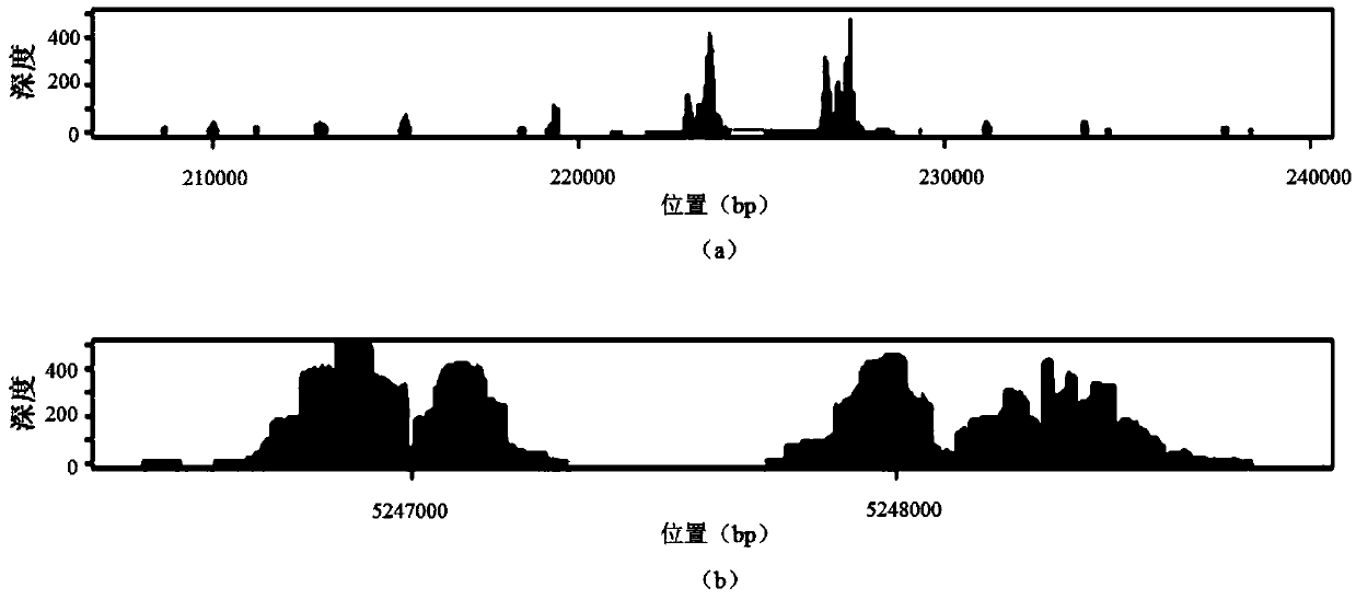
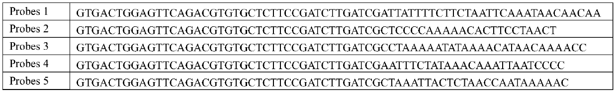
[0096] Step 1: Use magnetic beads or DSP Blood Mini kit (Qiagen) kit to extract free DNA in the isolated liver plasma, and analyze the extracted product by Agilent 2100 to detect the distribution of library fragments. It is found that the main peak of free DNA appears at 161bp, and then The multiple fragment distribution of 161bp appeared sequentially.

[0097] Step 2: Add 0.8μl T4DNA Kinase, 0.8μl T4DNA Polymerase, 0.2μl Klenow Polymerase, 1μl dNTP (10mM), 70mM Tris-HCl, 10mM MgCl to the PCR tube 2 , 5mM DTT, 0.2μl high temperature resistant polymerase, free DNA and sterile excess water, so that the total volume of the PCR reaction is 50μl. The PCR reaction conditions were 37°C for 30 minutes and 60°C for 30 minutes. After the reaction, add 1 μl of the first linker sequence shown in SEQ ID NO:4, 1 μl of the second linker sequence shown in SEQ ID NO:5, 8 μl of ATP (10 mM), 3 μl of T4 DNA ligase, 60 mM Tris-HCl (pH 7.6), ...

Embodiment 2

[0109] Example 2 Gene Mutation Detection

[0110] Step 1: Use magnetic beads or DSP Blood Mini kit (Qiagen) to extract genomic DNA from whole blood in vitro, fragment the genomic DNA to 100bp-1000bp by ultrasonic disruption, and elute the DNA to 30 μl of ultra- pure water.

[0111] Step 2: Add 0.8μl T4DNA Kinase, 0.8μl T4DNA Polymerase, 0.2μl Klenow Polymerase, 1μl dNTP (10mM), 70mM Tris-HCl, 10mM MgCl to the PCR tube 2, 5 mM DTT, 0.2 μl of thermostable polymerase and free DNA and sterile excess water to make a total reaction volume of 50 μl. The reaction conditions for end repair and addition of A were 37°C for 30 minutes and 50°C for 30 minutes. Add 1 μl of the first linker sequence shown in SEQ ID NO:4, 1 μl of the second linker sequence shown in SEQ ID NO:5, 8 μl of ATP (10 mM), 3 μl of T4 DNA ligase, 60 mM Tris-HCl ( pH 7.6), 10mM MgCl 2 , 1 mM ATP, 1 mM DTT and 7.5% PEG4000-8000, the total reaction volume was 80 μl. The linker ligation reaction conditions were 50 mi...


PUM


Abstract

The invention provides a detecting probe. The detecting probe comprises at least one capturing probe; sequences from the 5' end to the 3' end of the capturing probe sequentially include a preamble sequence, a probe random label sequence and a target complementary sequence; the preamble sequence is shown as SEQ ID NO:1; the probe random label sequence is composed of 0-12 random basic groups; and the target complementary sequence comprises at least 10 continuous basic groups complementary with a to-be-detected gene. The detecting probe is used for detecting multi-target-point gene mutation, methylation modification and/or hydroxymethylation modification, low in content requirement for the to-be-detected gene, capable of effectively avoiding disturbance of length and content of the to-be-detected gene to detection, beneficial to detection of genes with high degradability and serious fragmentation, low in detection cost, high in efficiency and wide in application range.

Description

technical field [0001] The invention relates to the field of medical biology, in particular to a detection probe, method and kit for detecting multi-target gene mutation, methylation modification and / or hydroxymethylation modification. Background technique [0002] The multi-site / region detection of genetic variation, methylation modification or hydroxymethylation modification of the target gene is of great scientific importance for the exploration of the molecular mechanism of disease occurrence, the screening and identification of molecular markers, assisting patient diagnosis and medication guidance. and clinical significance. In addition, establishing a genetic information database is also indispensable for analyzing and obtaining genetic information of rare or endangered samples. However, in most cases, the amount of DNA obtained from the sample is low and the fragments vary greatly, which is not conducive to the establishment of the library. For example, cfDNA (cell ...

Claims


Application Information

Patent Timeline
IPC IPC(8): C12N15/11C12Q1/6858
CPCC12Q1/6858C12Q2531/113C12Q2535/122C12Q2525/191C12Q2523/125
Inventor 王君文吉冠玉胡琪高飞
Owner SHENZHEN E GENE TECH
Why Eureka
Try Eureka