Opto-acoustic probe used for detecting nitroreductase in living bodies and preparation method and application thereof

A living body detection, reductase technology, applied in the field of tumor marker detection and imaging, to achieve the effect of accurate ratio detection, fast response speed, and large penetration depth

Inactive Publication Date: 2019-04-16
SOUTH CHINA NORMAL UNIVERSITY
View PDF2 Cites 9 Cited by
  • Summary
  • Abstract
  • Description
  • Claims
  • Application Information

AI Technical Summary

Problems solved by technology

[0005] The main disadvantage of this type of method is that in live detection, the scattering of ultrasonic signals by biological tissues to physiological tissues of the human bo

Method used


Image

  • Opto-acoustic probe used for detecting nitroreductase in living bodies and preparation method and application thereof
  • Opto-acoustic probe used for detecting nitroreductase in living bodies and preparation method and application thereof
  • Opto-acoustic probe used for detecting nitroreductase in living bodies and preparation method and application thereof

Examples


Embodiment 1

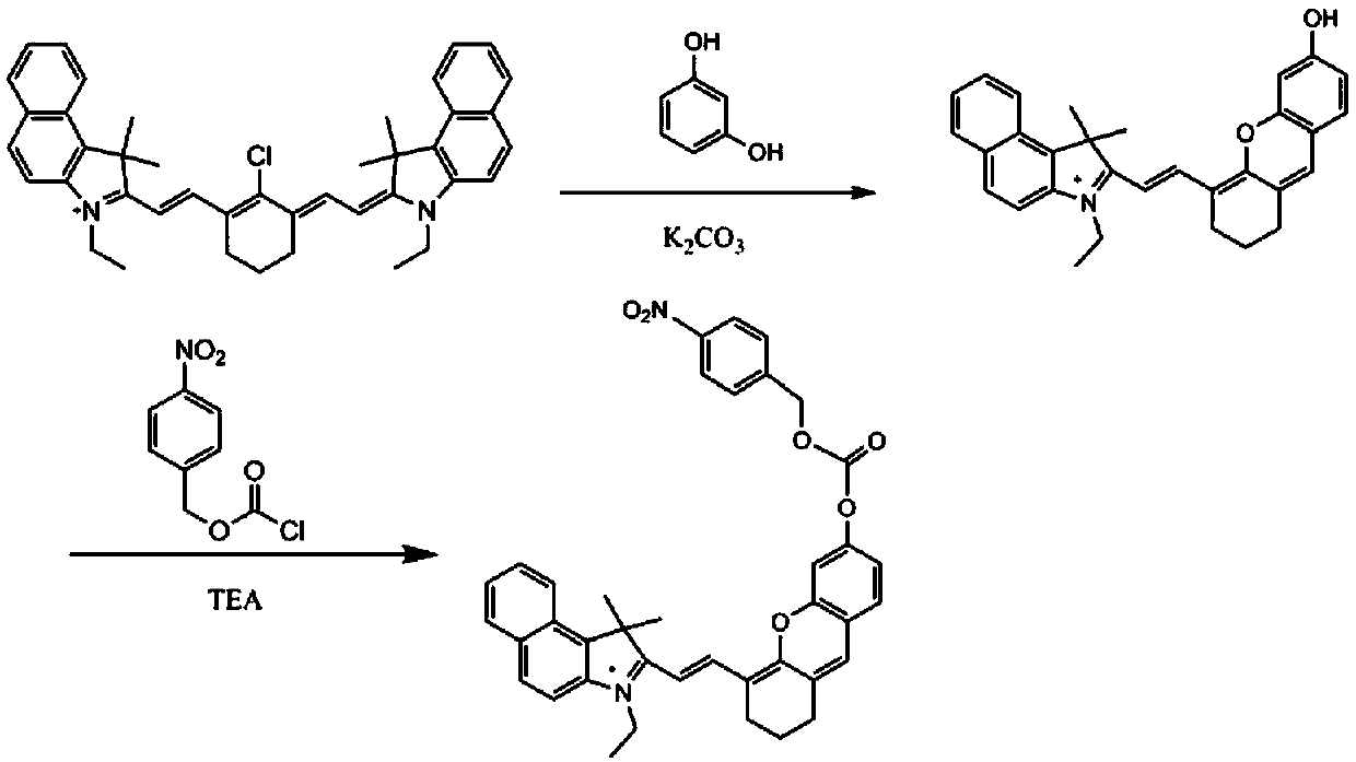
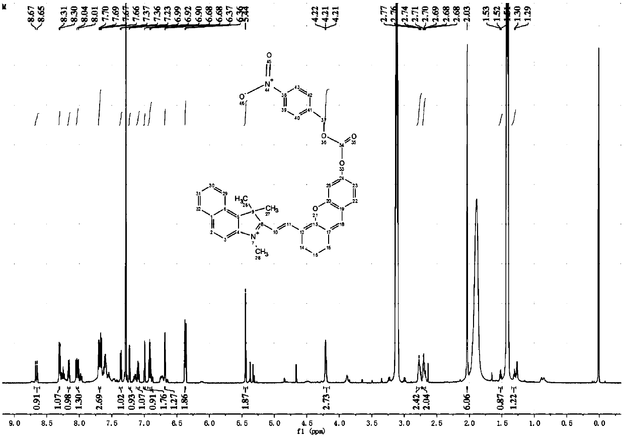
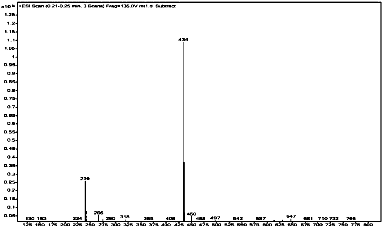
[0058] Example 1: Preparation of a photoacoustic probe for detecting nitroreductase in tumors in vivo.

[0059] The specific synthesis steps of a photoacoustic probe for in vivo detection of nitroreductase provided by the present invention are as follows: figure 1 As shown, the details are as follows:

[0060] ①Dissolve 20mL of frozen DMF in 20mL of dichloromethane under nitrogen protection to obtain a DMF solution;

[0061] ②Dissolve 10.5mL of phosphorus oxychloride in 10mL of dichloromethane under an ice bath, then add the solution of phosphorus oxychloride in dichloromethane dropwise into the DMF solution obtained in step ①, and stir for 30min under nitrogen protection to obtain mixture;

[0062] ③Add 5.26mL cyclohexanone to the mixed solution obtained in step ②, stir the mixture at 80°C, and reflux for 3h;

[0063] ④Then pour the mixture obtained in step ③ into ice water, overnight, and filter to obtain a yellow solid;

[0064] ⑤Dissolve 2mL of 1,2,2-trimethylbenzindol...

Embodiment 2

[0074] Example 2: Absorption before and after the reaction of probe I with nitroreductase.

[0075] Configure the probe I solution and the nitroreductase aqueous solution obtained in Example 1, then mix the probe I solution and the nitroreductase aqueous solution for five minutes and measure its absorption; wherein, the concentration of the probe I in the reaction system is 100 μM, the concentration of nitroreductase ranged from 0 to 50 μg / mL (0, 5, 10, 15, 20, 25, 30, 35, 40, 50 μg / mL). Absorption spectrum such as Figure 4 As shown, it can be seen from the figure that the absorption spectrum of the probe is red-shifted after reacting with nitroreductase.

Embodiment 3

[0076] Example 3: Photoacoustic imaging images of probe I reacted with different concentrations of nitroreductase.

[0077] A total of 6 groups of probe Ⅰ solutions with a concentration of 100 μM, nitroreductase aqueous solution with a concentration of 0, 10, 20, 30, 40, 50 μg / mL, and 5 mmol / L NADH solution were prepared, and then the probe Ⅰ solution was mixed with nitrate Reductase aqueous solution and NADH solution were mixed and reacted at 37°C (the amount of probe Ⅰ solution was 40 μL, the amount of nitroreductase aqueous solution was 1 μL, and the amount of NADH was 5 μL). After each group reacted for 5 minutes, use Photoacoustic computed tomography scanner measured photoacoustic signals of 6 groups of solutions, and photoacoustic two-dimensional images ( Figure 5 ). It can be seen from the figure that the intensity of the photoacoustic signal at 730 nm gradually increases with the increase of the concentration, indicating that the probe can be used as a ratiometric ph...


PUM


Abstract

The invention discloses an opto-acoustic probe used for detecting nitroreductase in living bodies and a preparation method and application thereof, and belongs to the field of detection and imaging oftumor markers. The structure formula of the opto-acoustic probe used for detecting nitroreductase in living bodies is shown in the formula I. The probe mainly takes benzo Cy7-C1 as a raw material, and through nucleophilic substitution, the crewengel condensation reaction and the chloroformate nucleophilic substitution reaction, the probe is obtained. The problems of scattering of a fluorescence signal and tissue absorption in the process of detection for the living bodies by adopting a fluorescence probe are solved, the prepared probe has the advantages that the penetration depth of an acoustical signal is large, imaging of deep tissue is achieved, response is quick, the specificity is good, and ratio detection is more accurate. The probe can be used for detecting the nitroreductase in the living bodies, thereby monitoring anaerobic conditions of tumors; the probe can also be applied to detection of the nitroreductase in cells and biological samples.

Description

technical field [0001] The invention belongs to the field of tumor marker detection and imaging, and particularly relates to a photoacoustic probe for detecting nitroreductase in a living body, a preparation method and application thereof. Background technique [0002] Now cancer has become the number one killer affecting human health, so research on cancer is the top priority in the work of current scientific researchers. Hypoxia is one of the hallmarks of solid tumors. Clinical studies have shown that hypoxia can block the metabolic process of many anticancer drugs, thereby affecting cancer. The efficacy of anticancer drugs, and hypoxia can promote the metastasis of tumor cells. In the research of cervical cancer and head and neck squamous carcinoma, hypoxia is even considered as a signal of progression-free survival in the detection, so the study of the hypoxia state of tumor cells has very important clinical significance (Kizaka-Kondoh, S.; Inoue, M.; Harada, H.; Hira...

Claims


Application Information

Patent Timeline
IPC IPC(8): C07D405/06G01N21/17
CPCC07D405/06G01N21/1702
Inventor 张涛邢达马腾
Owner SOUTH CHINA NORMAL UNIVERSITY
Why Eureka
Try Eureka