Preparation method of composite for treating brain glioma
A glioma and complex technology, which is applied in the directions of non-active ingredients medical preparations, medical preparations containing active ingredients, nano-drugs, etc., can solve the problem of low yield, inability to guarantee the purity of exosomes, and complex modification methods. and other problems to achieve the effect of high drug loading
- Summary
- Abstract
- Description
- Claims
- Application Information
AI Technical Summary
Problems solved by technology
Method used
Image

Examples
Embodiment 1
[0021] Embodiment 1: Preparation of complex (Exosome-DOX Nano)
[0022] 1. Preparation of complex (Exosome-DOX Nano)
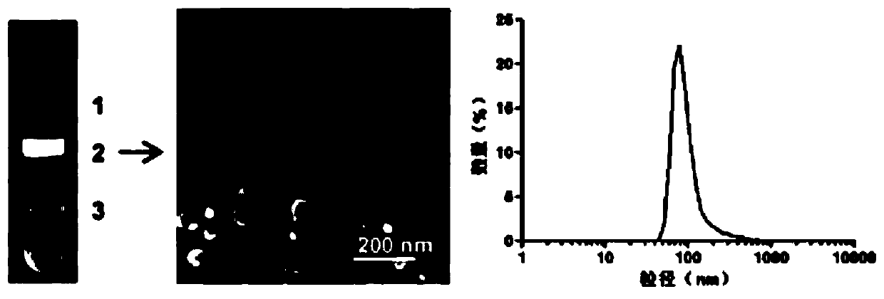
[0023] (1) Extraction and purification of exosomes: fresh grapefruit peeled and squeezed for juice, gradient centrifugation of grapefruit juice: 500g, 10min; Centrifuge for 1 h, take the precipitate, resuspend the precipitate with PBS buffer, and extract exosomes by sucrose density gradient centrifugation (8%, 30%, 45%, 60% sucrose-Tris.HCl solution), and use transmission electron microscopy and dynamic light scattering The appearance characteristics and particle size of exosomes were detected, and the concentration of exosomes was detected by BCA protein quantification method, and the obtained exosomes (Exosomes) were subpackaged and frozen at -80°C.
[0024] see figure 1 , transmission electron microscopy showed that the extracted exosomes were particles with a lipid bilayer structure and a spherical shape, and a dynamic light scattering instrument showed ...
Embodiment 2
[0035] Example 2. Composite (Exosome-DOX Nano) toxicity test on glioma cells
[0036] Resuscitate human glioma cells U87 and U251. In the logarithmic growth phase of the cells, inoculate the U87 and U251 cells in a 96-well plate with a cell number of about 3000 per well. Set 5 concentration gradients for the standard, that is, 0.2 μg / mL, 0.1 μg / mL, 0.05 μg / mL, 0.025 μg / mL, 0.0125 μg / mL, 0 μg / mL to act on the cells, and set 5 duplicate holes for each concentration. Routine culture in the dark for 48 hours after adding the drug, add MTT to continue the culture for 4 hours, detect the absorbance of each well at 490nm with a microplate reader, according to the formula relative cell viability (%)=(OD experimental group mean / OD control group mean)×100 % Calculate the relative survival rate of each group of cells respectively, the results are as follows Figure 6 As shown, the inhibitory ability of doxorubicin and doxorubicin nanomedicine to U87 is close, and the toxicity of the com...
Embodiment 3
[0037] Example 3. Distribution of compound exosomes in nude mice with orthotopic glioma implantation
[0038] When the nude mice grow to about 18 g, the nude mice are anesthetized with pentobarbital sodium, and 150,000 U87 glioma cells overexpressing luciferase are planted in the right striatum of the nude mouse brain. After about 3-4 days, the peritoneal The luciferase substrate was injected, and the growth of intracranial tumors in nude mice was observed 10 minutes later using a small animal in vivo imager. When the intracranial tumor grows to an appropriate size, a certain amount of cy7-labeled Exosome-cy7, DOX Nano-cy7, Exosome-DOX Nano-cy7 (the total amount of cy7 in each group is equal) is injected into the tail vein, and the nude mice are killed at 96 hours, and their hearts are collected. , liver, spleen, lung, kidney and brain, respectively observe the distribution of drugs in each organ and the localization of drugs in brain tumors, the results are as follows Figur...
PUM
| Property | Measurement | Unit |
|---|---|---|
| The average particle size | aaaaa | aaaaa |
| The average particle size | aaaaa | aaaaa |
Abstract
Description
Claims
Application Information