
Preparation method and application of endothelial vesicles of Escherichia coli with endogenous high expression of microRNA in anti-tumor drug preparation

A technology of anti-tumor drugs and Escherichia coli, which is applied in the direction of anti-tumor drugs, biochemical equipment and methods, DNA/RNA fragments, etc., can solve the problems that have not been seen in the application of drug-loaded intimal vesicles, and achieve growth inhibition, The effect of simple operation and low production cost

Active Publication Date: 2019-07-23
ZHENGZHOU UNIV
View PDF4 Cites 3 Cited by
  • Summary
  • Abstract
  • Description
  • Claims
  • Application Information

AI Technical Summary

Problems solved by technology

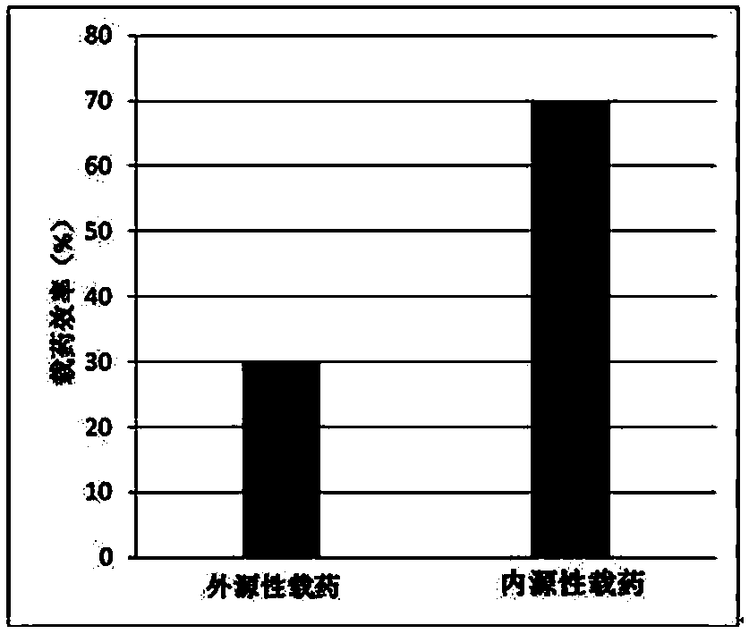
Although these new technologies have improved the efficiency of drug delivery to a certain extent, simplified the operation process, and got rid of the dependence on expensive equipment and facilities, they are still limited to the technical level of extracellular drug loading. Application of drug loading in membrane vesicles

Method used


Image

  • Preparation method and application of endothelial vesicles of Escherichia coli with endogenous high expression of microRNA in anti-tumor drug preparation
  • Preparation method and application of endothelial vesicles of Escherichia coli with endogenous high expression of microRNA in anti-tumor drug preparation
  • Preparation method and application of endothelial vesicles of Escherichia coli with endogenous high expression of microRNA in anti-tumor drug preparation

Examples


Embodiment 1

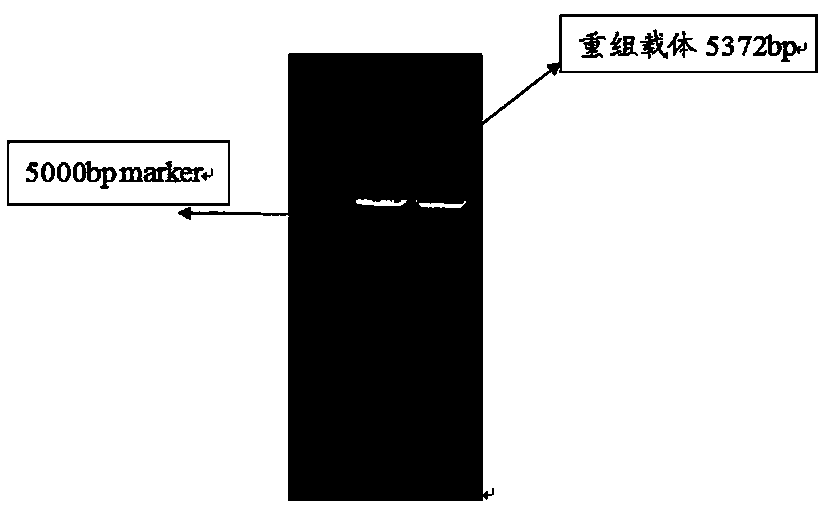
[0034] Example 1 Preparation of Escherichia coli endogenously expressing miRNA in large quantities

[0035] Step 1, design the primer sequence of pET-31b(+) and PCR amplify the whole gene sequence of pET-31b(+)

[0036] (1) Design the primer sequence of pET-31b(+) vector

[0037] The sequence is as follows:

[0038] Upstream primer: 5'-AAGAATTCAAGCTTATTCTCTTCTTAAAGTTAAACAAAATTATT-3';

[0039] Downstream primer: 3'-TTGGATCCGTCGAC GCAATAACTAGCATAACCCCTTG-5'.

[0040] (2) Extract the plasmid of K12 Escherichia coli as a template for PCR amplification of pET-31b(+) vector

[0041] a. Take out the K12 Escherichia coli strain preserved in 20% glycerol from the -80 low-temperature refrigerator, wait for it to melt, and streak it on a solid LB plate containing 100 mg / ml ampicillin in an ultra-clean workbench, and invert at 37°C Cultivate for 12-16 hours;

[0042] b. Pick a single clone in 5ml LB liquid medium containing 100mg / ml ampicillin, and shake and culture at 37°C and 220rm...

Embodiment 2

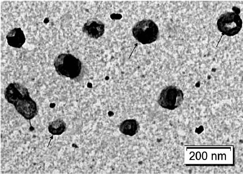
[0115] Example 2 Preparation of Escherichia coli inner membrane vesicles

[0116] 1. Use lysozyme to remove the outer cell membrane of E. coli to obtain E. coli protoplasts

[0117] a. Prepare 50mM Tris-HCl (pH 8.0), 50mM Glucose, and 1mM EDTA solution respectively as a buffer solution, and prepare lysozyme with a concentration of 4mg / ml;

[0118] b. Add lysozyme to the collected Escherichia coli cells (prepared in Example 1), react in a 37°C water bath for 20 minutes, and remove toxins such as the outer membrane of the cell and lipopolysaccharide on it;

[0119] c. Take out a little bacterial liquid and observe and detect under a microscope until completely de-walled protoplasts are obtained.

[0120] 2. Use a polycarbonate membrane to filter E. coli protoplasts to obtain nanovesicles in the inner membrane of E. coli protoplasts

[0121] a. Install a polycarbonate membrane with a pore size of 12um on a filter with a size of 47mm to filter E. coli protoplasts;

[0122] b. I...

Embodiment 3

[0125] Example 3 Fluorescent quantitative PCR detection of miR-34a in Escherichia coli inner membrane vesicles prepared in Example 2

[0126] a. Use the method provided in Example 2 to isolate E. coli inner membrane vesicles, resuspend them with 100ul PBS, use 500ul RLSolution to fully lyse the bacteria, add 40ul chloroform and mix well, centrifuge at 12000rpm for 10min, and carefully pipette 200ul of the supernatant to fresh Add 400ul of absolute ethanol to a centrifuge tube, mix thoroughly and add to the chromatography column, centrifuge at 12000rpm for 1 minute, pour off the filtrate, wash twice with Wash Buffer, and finally use RNase-free DEPC water to elute RNA to obtain fresh RNA;

[0127] b. Use the downstream primer of miR-34a in step 2 for reverse transcription, and reverse transcribe the RNA obtained in the previous step into cDNA; RNA 3ul, 10×Buffer 2ul, reverse transcriptase 2ul, downstream primer 1ul, DEPC water 12ul, the total system is 20ul, select the reverse ...


PUM


Abstract

The invention discloses a preparation method of endothelial vesicles of Escherichia coli for anti-tumor drugs with endogenous high expression of nucleic acids. According to the preparation method of the endothelial vesicles of Escherichia coli, a 'tRNA scaffolding means' is adopted so as to stably insert microRNA into a vector; the vector is transformed into Escherichia coli, thereby allowing highendogenous expression of microRNA with cancer-cell-killing effects; epicellular and periplasmic components of the Escherichia coli are removed by using lysozyme, thereby obtaining protoplast of Escherichia coli; the protoplast is filtered by using polycarbonate membrane, thereby having the protoplast broken; and then, purification is carried out by adopting a ultracentrifugation means so as to have endothelial vesicles of the protoplast separated, thereby obtaining the endothelial vesicles of Escherichia coli with high expression of miRNA and low toxicity. The preparation method provided by the invention is easy to operate, low in production cost, feasible for large-scale fermentation and preparation, low in toxicity, and highly efficient. As a novel pharmaceutical carrier, being appliedin anti-tumor drug preparation, the endothelial vesicles of Escherichia coli are capable of significantly inhibiting development of non-small cell lung cancer; and thus, the endothelial vesicles of Escherichia coli have broad application prospects in the field of pharmaceutical carriers.

Description

technical field [0001] The present invention relates to biotechnology, in particular to a method for preparing endogenous endogenous high-expression miRNA low-endotoxin Escherichia coli intimal vesicles, and the present invention also relates to the use of prepared intimal vesicles in the preparation of antitumor drugs application. Background technique [0002] The method of using bacterial nanovesicles to load drugs has gradually attracted people's attention in recent years because of its low cost of mass production, easy modification, strong stability, and relatively simple surface proteins, which can greatly reduce immunogenicity after modification. . Gram-negative bacteria have two membranes—an outer membrane and an inner membrane. The outer membrane contains toxins such as lipopolysaccharides, while the inner membrane contains fewer toxins than the outer membrane. The study found that both the outer and inner membranes of Gram-negative bacteria can produce nano-sized ...

Claims


Application Information

Patent Timeline
IPC IPC(8): A61K9/50A61K31/7105A61K47/46A61P35/00C12N15/113C12N15/70
CPCC12N15/70C12N15/113A61K9/5068A61K31/7105A61P35/00C12N2310/141
Inventor 翁海波崔晨阳郭婷婷张帅
Owner ZHENGZHOU UNIV
Learn More