Method for constructing tissue engineered cartilage by using bone-marrow mesenchymal stem cells

A bone marrow mesenchymal and tissue engineering technology, applied in the direction of bone/connective tissue cells, animal cells, vertebrate cells, etc., can solve the problems of bone marrow mesenchymal stem cells new tissue engineering cartilage vacuolization and other problems, and achieve central vacuolation. The effect of chemical suppression and simple operation

Active Publication Date: 2019-08-27
FOURTH MILITARY MEDICAL UNIVERSITY
View PDF4 Cites 1 Cited by
  • Summary
  • Abstract
  • Description
  • Claims
  • Application Information

AI Technical Summary

Problems solved by technology

However, these techniques usually require the use of a large amount of exogenous growth factors and a large number of BMSC in vitro culture operations before in vivo culture. At the same time, how to inhibit the hypertrophy and ossification of bone marrow mesenchymal stem cells in vivo and how to prevent new tissue engineering cartilage Cavitation still needs to be solved

Method used


Image

  • Method for constructing tissue engineered cartilage by using bone-marrow mesenchymal stem cells
  • Method for constructing tissue engineered cartilage by using bone-marrow mesenchymal stem cells
  • Method for constructing tissue engineered cartilage by using bone-marrow mesenchymal stem cells

Examples


Embodiment 1

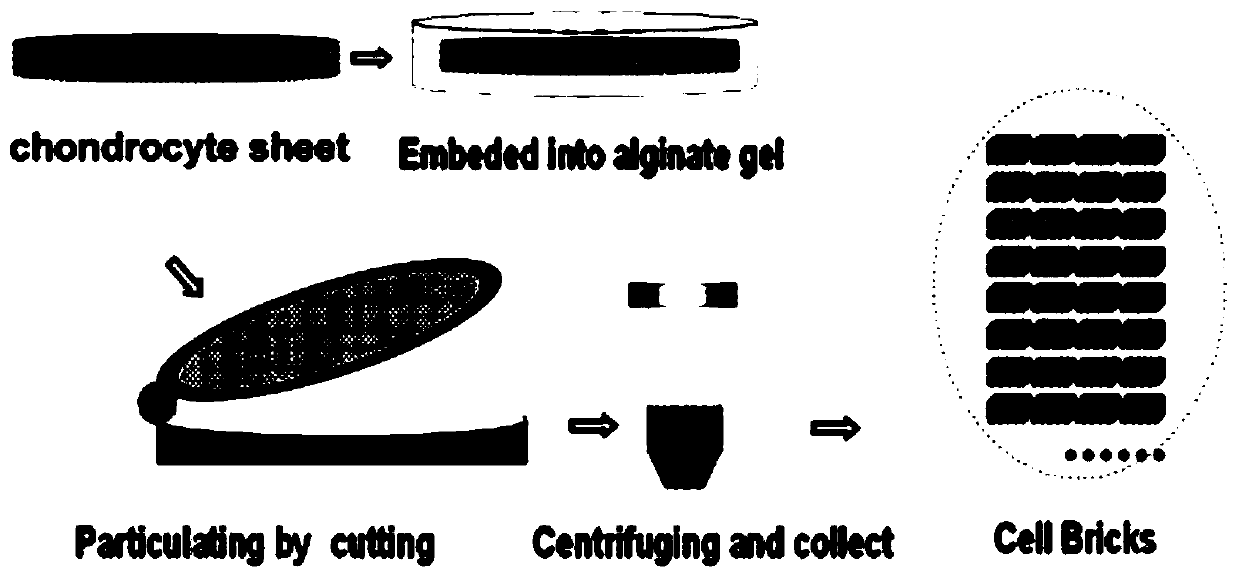
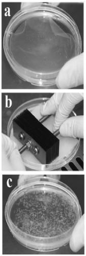
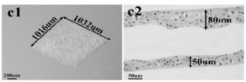
[0025] Example 1. Preparation of chondrocyte aggregates (cell bricks)

[0026] Take the ear cartilage of 1-month-old New Zealand white rabbit, cut it into pieces, digest it with 0.2% (wt) type II collagenase (Gibco, USA) solution at 37°C for 12 hours, and then take the single cell suspension in a 15mL centrifuge tube Centrifuge at 1000rpm for 5min. The supernatant was discarded, washed once with PBS, centrifuged at 1000 rpm for 5 min, and the cells were resuspended in membrane-forming inducing solution. Take 6.5×10 5 cells / cm 2 The concentrations were grown in 6-well plates. Afterwards, the film-forming inducing medium was used to continue culturing, and the medium was changed every 3 days. The film-forming induction medium used was high-glucose DMEM medium (Hyclone, U.S.), containing 20% ​​fetal bovine serum (Gibco, U.S.), 272 μg / mL glutamine (Amresco, U.S.), 50 μg / mL ascorbic acid (Amresco, U.S. ), 50 μg / mL penicillin (Amresco, USA), 30 μg / mL penicillin (Amresco, USA)...

Embodiment 2

[0029] Example 2. Preparation of platelet-rich plasma (PRP)

[0030] Take 2-month-old New Zealand white rabbit ear vein blood, add 3.8% (wt) sodium citrate solution anticoagulant according to 10% of the total volume, and prepare PRP by two-step centrifugation: whole blood is centrifuged at 1800rpm at room temperature After 8 minutes, divide into upper, middle and lower layers, the upper layer is platelet-poor plasma, the middle layer is platelet-rich plasma, and the lower layer is red blood cells; transfer the upper and middle layers of plasma to another centrifuge tube, centrifuge at 3600rpm for 8 minutes, discard the upper layer 3 / 4 of the platelet-poor plasma, and the rest of the platelets were blown repeatedly with a dropper to form platelet-rich plasma, in which the final concentration of platelets was 20.9±1.1×10 8 cells / mL and kept on ice for future use.

Embodiment 3

[0031] Example 3. Preparation, in vivo growth and detection results of BMSC-cell brick-platelet cell complexes

[0032] Experimental operation:

[0033] Suspend the BMSC subcultured to the P3 generation and the cell brick (CB) prepared in Example 1 in 500 μL of the platelet-rich plasma prepared in Example 2, mix well, and add 50 μL of thrombin solution containing 100 mg / mL calcium chloride to it . Then injected subcutaneously into nude mice for in vivo growth. In this experiment, we focused on the effect of the ratio between the number of BMSCs and the number of chondrocytes contained in cell bricks on the formation of nascent tissue-engineered cartilage. The sum of the number of BMSCs and chondrocytes used was 3 × 10 8 indivual. The experiments performed were divided into three groups, including ratios between BMSCs and chondrocytes of 1:2, 1:1, and 2:1, hereinafter denoted as B1CB2, B1CB1, and B2BC1, respectively. After 8 weeks of in vivo growth, the samples were take...


PUM

PropertyMeasurementUnit
areaaaaaaaaaaa
thicknessaaaaaaaaaa

Abstract

The invention provides a method for constructing tissue engineered cartilage by using bone-marrow mesenchymal stem cells, which comprises the steps of (1) preparing a chondrocyte aggregate, (2) centrifuging whole blood to prepare platelet-rich plasma, (3) placing the bone-marrow mesenchymal stem cells and the chondrocyte aggregates in the platelet-rich plasma, uniformly mixing, and adding a thrombin solution containing calcium chloride to the obtained mixture to form a cell complex; (4) injecting the cell complex into a subject for growth in vivo to obtain the tissue engineered cartilage, wherein the number ratio of the bone mesenchymal stem cell to chondrocyte contained in the chondrocyte aggregate in step (3) is 1:1. The method for constructing tissue engineered cartilage of the invention is simple in operation, can maintain optimal new cartilage morphology and cartilage differentiation characteristics, and effectively inhibits the occurrence of new cartilage edge ossification whileeffectively resisting central vacuolization.

Description

technical field [0001] The invention relates to a method for constructing tissue engineered cartilage, in particular to a method for constructing tissue engineered cartilage by using bone marrow mesenchymal stem cells (BMSC) and chondrocytes. Background technique [0002] Currently, bone marrow mesenchymal stem cells are used in some techniques for constructing tissue engineered cartilage. Bone marrow mesenchymal stem cells have the potential of chondrogenic differentiation. When they are co-cultured with chondrocytes, the microenvironment composed of chondrocytes and their secreted cytokines is conducive to the chondrogenic differentiation of BMSCs. However, these techniques usually require the use of a large amount of exogenous growth factors and a large number of BMSC in vitro culture operations before in vivo culture. At the same time, how to inhibit the hypertrophy and ossification of bone marrow mesenchymal stem cells in vivo and how to prevent new tissue engineering c...

Claims


Application Information

Patent Timeline
Patent Type & Authority Applications(China)
IPC IPC(8): C12N5/077C12N5/0775
CPCC12N5/0663C12N5/0655
Inventor 巴睿恺赵铱民吴炜
Owner FOURTH MILITARY MEDICAL UNIVERSITY
Why Eureka
Try Eureka