A zebrafish brain trauma model and its preparation method and application
A zebrafish and brain trauma technology, applied in the field of zebrafish brain trauma model and its preparation, can solve problems such as non-traumatic brain trauma model, achieve good prospects and value, and promote the effect of repair
- Summary
- Abstract
- Description
- Claims
- Application Information
AI Technical Summary
Problems solved by technology
Method used
Image
Examples
Embodiment 1
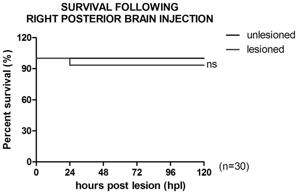
[0029] Example 1 Construction of Zebrafish Brain Injury Model
[0030] 1. Experimental method
[0031] 1. Zebrafish rearing and embryo reproduction
[0032] Commercially available enhanced green fluorescent protein-labeled macrophages (microglia distributed in the brain, hereinafter referred to as microglia) transgenic zebrafish (coro1α: EGFP zebrafish) were used as research objects; The Eisen zebrafish breeding system was used to feed brine shrimp twice a day, with 14 hours of light and 10 hours of darkness. The temperature is 28.5°C, the pH is 7.0-8.0, the dissolved oxygen value is 4.5-5.5 mg / l, and the conductivity is 550-650 μS / cm.
[0033] Coro1α: EGFP zebrafish were mated, male fish: female fish = 1: (1~2).
[0034] Embryos were collected and cultured in an embryo incubator at 28.5°C. Dead eggs were picked every day, embryo water was changed, and embryo water containing 0.003% (g / ml) PTU was changed at 1 dpf (days post fertilization) to inhibit pigment production. At...
Embodiment 2
[0051] Example 2 In situ hybridization method to detect the changes of inflammatory factors at different time points
[0052]1. Experimental method
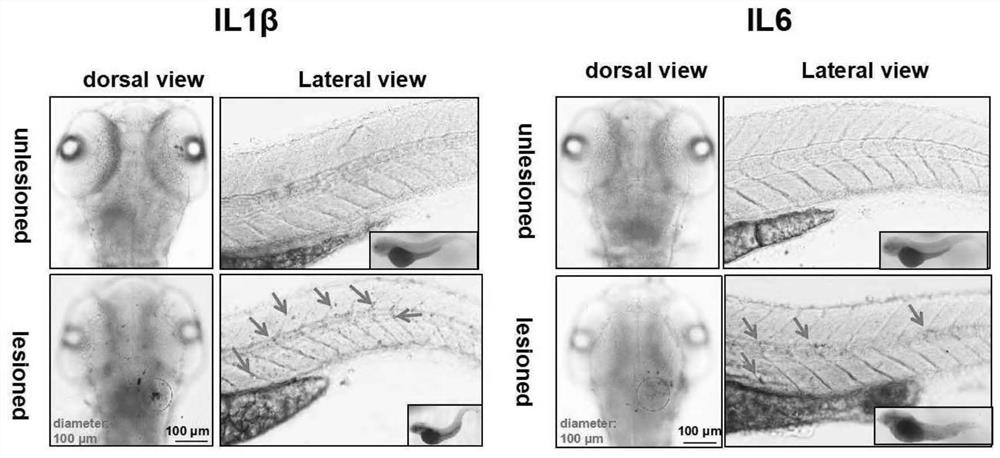
[0053] Taking the animal model constructed in Example 1 as the object (trauma group), with untreated zebrafish of the same age as the control (non-trauma group), the embryos of 1hpl and 24hpl after the trauma were collected for the detection of IL1-β and IL-6 inflammatory factors. in situ hybridization. 30 embryos of 1 hpl (hourspost lesion, 1 hour after wounding) were collected from the non-trauma group (untreated zebrafish group of the same age) and the wound group (animal model constructed in Example 1) respectively, and fixed overnight at 4° C. with 4% PFA. The temporal and spatial changes of the transcriptional levels of inflammatory factors IL1-β and IL-6 were detected by in situ hybridization. Probe concentration: IL1-β: 2.4ng / μl, IL-6: 3.0ng / μl.
[0054] 2. Experimental results
[0055] see results figure 2 , IL1-β ...
Embodiment 3
[0056] Example 3 Real-time tracking of changes in microglia at the trauma site after intervention
[0057] 1. Experimental method
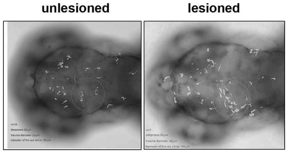
[0058] Taking the animal model constructed in Example 1 as the object (injury group) and untreated zebrafish of the same age as the control (non-injury group), confocal tracking of the aggregation of microglia at the wound site at 1hpl and 24hpl.
[0059] Video: Using FV3000 Olympus Confocal, the setting parameters are: Format: 1024×1024, Nr.ofsteps: 15, Z-step size: 2.92μm, Z-Volume: 40.804μm, Time Interval: 1min, Duration: 1h , stacks: 60.
[0060] Photographing: Use confocal to collect 1hpl and 24hpl images of non-trauma group and trauma group, setting parameters: Format: 1024×1024, Nr.of steps: 15, Z-step size: 2.92μm, Z-Volume: 40.804μm.
[0061] 2. Experimental results
[0062] Such as image 3 As shown, there was no microglial cell aggregation within 160 μm around the trauma site in the non-trauma group, but there was obvious microglial...
PUM
 Abstract
Description
Claims
Application Information
 - R&D
- Intellectual Property
- Life Sciences
- Materials
- Tech Scout
- Unparalleled Data Quality
- Higher Quality Content
- 60% Fewer Hallucinations
Browse by: Latest US Patents, China's latest patents, Technical Efficacy Thesaurus, Application Domain, Technology Topic, Popular Technical Reports.
© 2025 PatSnap. All rights reserved.Legal|Privacy policy|Modern Slavery Act Transparency Statement|Sitemap|About US| Contact US: help@patsnap.com


