A preparation method of self-assembled prevascularized stem cell membrane

A cell membrane, pre-vascular technology, applied in medical science, prosthesis, etc., can solve the problems of cell necrosis, unable to form tissue regeneration effect, etc.

Active Publication Date: 2020-10-23
FOURTH MILITARY MEDICAL UNIVERSITY
View PDF4 Cites 0 Cited by
  • Summary
  • Abstract
  • Description
  • Claims
  • Application Information

AI Technical Summary

Problems solved by technology

When using a multi-layer cell membrane to repair tissue, the diffusion effect can only ensure the survival of the cells in the surface layer, while the cells in the deep inner layer often suffer from necrosis due to lack of nutrients, thus failing to form a good tissue regeneration effect

Method used


Image

  • A preparation method of self-assembled prevascularized stem cell membrane
  • A preparation method of self-assembled prevascularized stem cell membrane
  • A preparation method of self-assembled prevascularized stem cell membrane

Examples


Embodiment 1

[0027] Example 1 Construction of vascularized EC-BMSC membrane

[0028] 1. Isolation, culture and identification of hBMSC

[0029] Use the whole bone marrow adherent method to isolate and culture hBMSC: Take fresh aseptic bone marrow, add an equal amount of PBS to mix, centrifuge at 800 rpm for 5 minutes, discard the supernatant, wash three times, add 15% fetal bovine serum and 0.4mg / mL of glutamine-based α-MEM medium, inoculated in 75 cm 2 in a culture bottle. After 3 days, half of the medium was changed for the first time, and then a new medium was replaced the next day to remove unadhered cells, and the adherent cells were continued to be cultured. The medium was changed every 2 days. When the cells grew to cover about 80% of the culture flask, they were digested with 0.25% trypsin and subcultured. Cells of passage P4-P5 were selected for subsequent operations.

[0030] 2. Isolation and culture of hUVEC

[0031] Take a sterile free umbilical cord, separate the umbilic...

Embodiment 2

[0040] Example 2 In vitro and in vivo functional evaluation of vascularized EC-BMSC composite membrane

[0041] 1. Construction of vascularized EC-BMSC composite membrane

[0042] The construction of the vascularized EC-BMSC composite membrane is the same as that in Example 1.

[0043] 2. Protein microarray analysis of supernatant of co-cultured cells

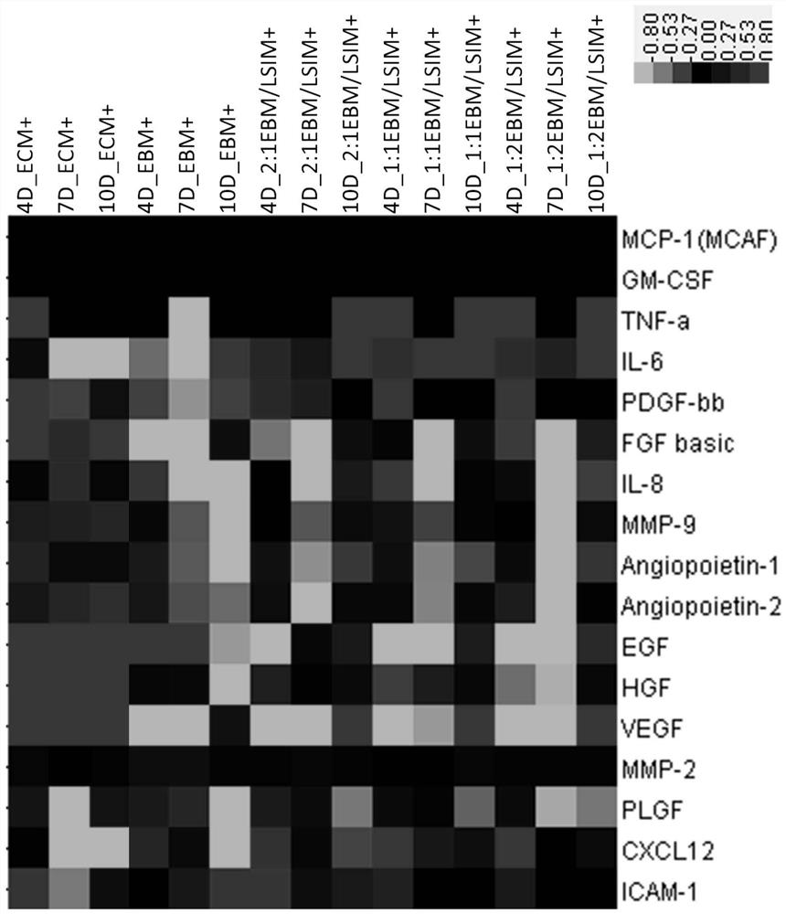
[0044] The cell supernatants of the co-culture system at different time points (3 days, 7 days, 10 days) were collected, and protein microarrays were used to analyze the differences in the expression of related secretory proteins (Table 2) in the co-culture supernatants. The results are as follows image 3 Shown: EGF, HGF, FGF, VEGF, Ang-1, Ang-2, MMP-9, IL-8 were highly expressed in the supernatant of group 1# added with exogenous growth factors. On the one hand, the high content of exogenous VEGF factors will transform vascular endothelial cells into a large number of tip cells, resulting in excessive sprouting of blood ves...


PUM


Abstract

The invention discloses a method for preparing a self-assembled pre-vascularized stem cell membrane in vitro by utilizing paracrine effect of mesenchymal stem cells. The human mesenchymal stem cells (hMSC) are used to induce membrane formation in vitro for 6 days, MSC membranes are initially formed, then endothelial cells (EC) are directly inoculated on the surface at a saturation density, a nutrient solution is changed to a culture medium having co-culture condition for continuous culture for 10 days, and the construction of the pre-vascularized EC-MSC composite membrane is completed. Compared with the traditional method of adding growth factor to promote vascularization of the membrane, the vascular network formed by the vascularized membrane of the invention is more in conformity with physiological morphology and high blood vessel maturity, the preparation cost is low, and anastomosis is quickly formed with host vessel after implantation in vivo with no leakage. The technology can reconstruct a vascular system of the defect tissue in a short time, and contributes to the repair and regeneration of large-area tissue defects, which provides a new idea for vascular tissue engineering and has broad application value.

Description

technical field [0001] The invention belongs to the technical field of tissue engineering, in particular to a method for preparing a self-assembled prevascularized stem cell membrane. Background technique [0002] Cell sheet technology is to continuously culture high-density seeded cells in vitro, make them grow in layers, form a film-like structure rich in cells and extracellular matrix, and harvest them in a non-enzymatic way, which has a unique Preservation of cell-extracellular matrix and intercellular signaling molecules, high cell utilization, good plasticity and other advantages. This technology has become an important part of modern regenerative medicine and shows great prospects for clinical application. When using a multi-layered cell membrane to repair tissue, the diffusion effect can only ensure the survival of the cells in the surface layer, while the cells in the inner deep layer are often necrotic due to lack of nutrients, thus failing to form a good tissue r...

Claims


Application Information

Patent Timeline
Patent Type & Authority Patents(China)
IPC IPC(8): A61L27/38
CPCA61L27/3808A61L27/3834
Inventor 闫钧赵领洲胡祥翔吴赬
Owner FOURTH MILITARY MEDICAL UNIVERSITY
Why Eureka
Try Eureka