
Probe and application thereof

A probe and light reaction technology, applied in the biological field, can solve the problem of low efficiency of signal transduction complexes, and achieve the effect of effective extraction and enrichment

Active Publication Date: 2019-09-13
SOUTH UNIVERSITY OF SCIENCE AND TECHNOLOGY OF CHINA
View PDF8 Cites 5 Cited by
  • Summary
  • Abstract
  • Description
  • Claims
  • Application Information

AI Technical Summary

Problems solved by technology

Inefficient identification of tyrosine phosphorylated protein-mediated signaling complexes from complex biological samples using these methods

Method used


Image

  • Probe and application thereof
  • Probe and application thereof
  • Probe and application thereof

Examples


Embodiment 1

[0093] Embodiment 1 Probe backbone 2 labels SH2 bait protein

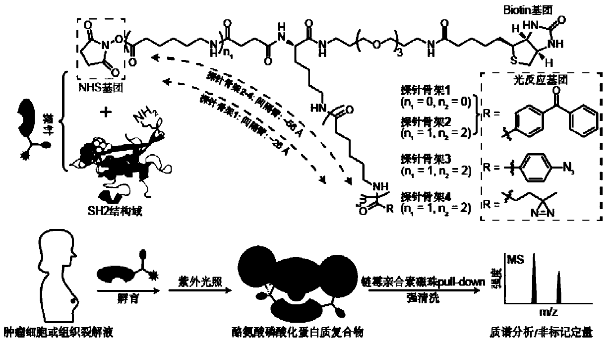
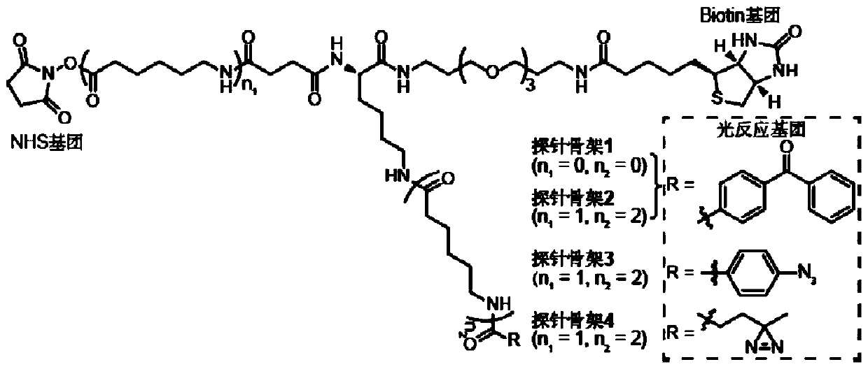
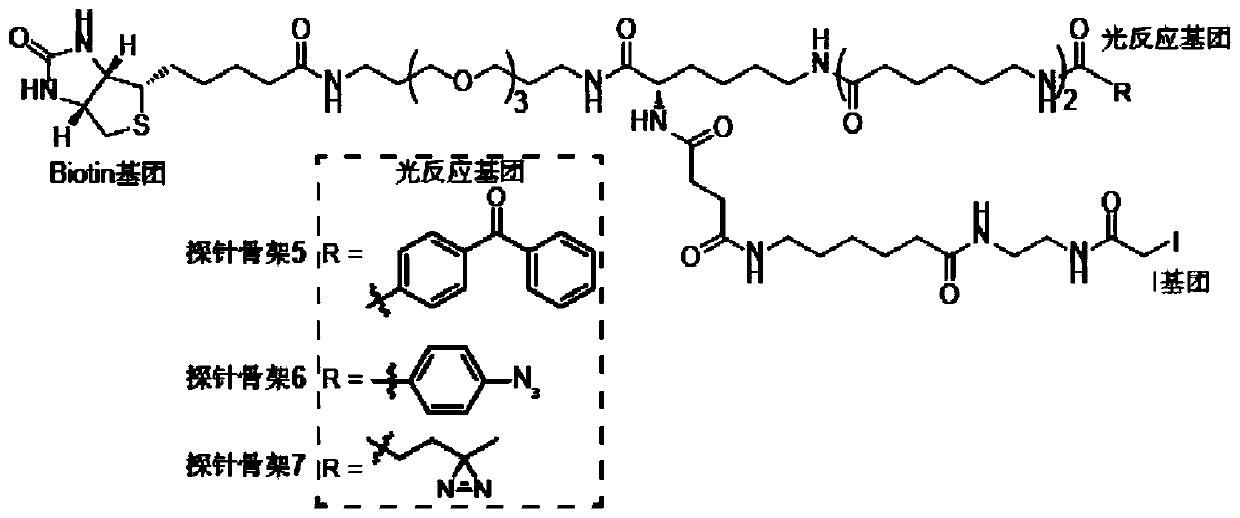
[0094] The present invention designs 7 kinds of probe skeletons, which are used to prepare such as Figure 1A The functional probe shown has high-efficiency enrichment and identification of tyrosine phosphorylated protein complexes. Among them, the structure of the probe skeleton is as follows Figure 1B with Figure 1C shown.

[0095] To 50 μL SH2 domain (1 mg / mL, dissolved in HEPES buffer), add 0.5 μL probe backbone containing photoreactive groups and biotin groups (probe backbone dissolved in DMSO, SH2 domain and The molar ratio of the probe skeleton is 1:10), reacted at room temperature for 1 min, then added 5 μL of 1 mol / L glycine solution to terminate the activity of the NHS group, and obtained the following Figure 1A Probes labeled with the SH2 domain of the bait protein are shown.

Embodiment 2

[0096] Example 2 Mass spectrometry sample pretreatment

[0097] (1) According to the method in Example 1, using probe skeletons 1-7 to prepare probes with different spacer arm lengths or photoreactive groups, add 1.4 mg of cell lysate, incubate slowly at 4 °C for 2 h, and then 365 nm ultraviolet light Light for 30min;

[0098] (2) Add 30 μL of streptavidin microspheres, incubate slowly at 4°C for 2 h, wash with 1 mL of mRIPA buffer three times, and wash once with 1 mL of 50 mM ammonium bicarbonate (ABC).

Embodiment 3

[0099] Embodiment 3 prepares mass spectrometry detection sample

[0100] (1) Incubation and digestion:

[0101] Add Tris(2-carboxyethyl)phosphine hydrochloride (Tris(2-carboxyethyl)phosphine hydrochloride, TCEP, dissolved in 50mM ABC) with a final concentration of 10mM to the sample pretreated in Example 2, and incubate at room temperature for 15min; Iodoacetamide (iodoacetamide, IAA, dissolved in 50 mM ABC) with a final concentration of 30 mM was incubated at room temperature for 15 min in the dark; TCEP with a final concentration of 5 mM was added and incubated at room temperature for 15 min; the sample was washed once with 1 mL of 50 mM ABC, and the Add 50 μL of trypsin solution (trypsin, 2 μg, dissolved in 50 mM ABC), and enzymatically digest at 37 ° C;

[0102] (2) Enzymatic peptide desalination:

[0103] 1) Collect the enzymolyzed polypeptide: centrifuge to collect the above-mentioned overnight enzymatically hydrolyzed sample, add 50 μL of 1% (v / v) formic acid, wash th...


PUM

PropertyMeasurementUnit
Wavelengthaaaaaaaaaa

Abstract

The present invention provides a probe and the application thereof. The probe comprises a spacer arm, and bait protein, a photoreactive group and an enriched group attached to the spacer arm. The baitprotein comprises a protein structure domain for identifying specific protein after translational modification, such as an SH2 domain for identifying tyrosine phosphorylation. According to the probeprovided by the present invention, the activity of the target protein cannot be affected by the probe, when the bait protein interacts with the target protein, ultraviolet radiation promotes covalentbinding of the photoreactive group and the target protein, and protein complexes are effectively enriched and obtained through the enriched group, so that analysis on weak interactions and transient interactions between proteins can be facilitated, enrichment and identification abilities of target proteins and complexes thereof in the human or animal tissue samples and near the cell membranes aresignificantly improved, enrichment and identification of tyrosine phosphorylation protein complexes in different breast cancer cell lines and breast cancer tissues are successfully realized, and the probe has broad application prospects and market value.

Description

technical field [0001] The invention belongs to the field of biotechnology, relates to a probe and its application, in particular to a probe containing three functional groups and its application in high-throughput detection of protein interaction. Background technique [0002] Protein is the main component of organisms. In organisms, hundreds of thousands of protein-protein interactions occur every moment. These huge interactions assemble proteins into a wide variety of protein complexes with different functions. , Execute and regulate almost all life processes and functions, including protein translation, cell cycle control, protein synthesis and degradation, etc. Protein complexes form a high-dimensional and complex cellular signal transduction network through serial and parallel interactions between each other; macroscopic and precise control of cellular functions is achieved through exogenous signal stimulation and precise positioning based on time and subcellular organ...

Claims


Application Information

Patent Timeline
IPC IPC(8): G01N33/68
CPCG01N33/68
Inventor 田瑞军李鹏飞初碧珠何岸郑振东
Owner SOUTH UNIVERSITY OF SCIENCE AND TECHNOLOGY OF CHINA
Learn More