

Hybridoma cell strain, getah virus distinguishing monoclonal antibody secreted from hybridoma cell strain, and application
A hybridoma cell line and monoclonal antibody technology, which is applied in the field of bioengineering, can solve the problems of preparation and application of GETV monoclonal antibody not seen in Gaita virus, and achieve the effects of wide application space, strong specificity and high sensitivity
- Summary
- Abstract
- Description
- Claims
- Application Information
AI Technical Summary
Problems solved by technology
Method used
Image

Examples
Embodiment 1
[0033] The preparation method of the anti-Gaita virus monoclonal antibody of the present invention comprises the following steps:
[0034] 1. Construction of E2 protein recombinant expression vector
[0035] Gatavirus RNA was extracted and reverse transcribed into cDNA. For specific operation steps, see the RNA extraction manual in the Trizol Invitrogen kit;
[0036] Take 300 μL of the virus solution and extract total virus RNA according to the instructions of TRIZOL RNA Extraction Reagent.
[0037] Reverse transcription reaction system (20 μL): RNA template 13 μL, 5×buffer 4 μL, dNTP (10 mmol / μL) 1 μL, random primer Random (20 pmol / μL) 1 μL, RNase inhibitor (40 U / μL) 0.5 μL, reverse Recorder M-MLV (200U / μL) 0.5 μL; reaction parameters: 42°C for 1h, 95°C for 5min, the cDNA product was used for PCR amplification.
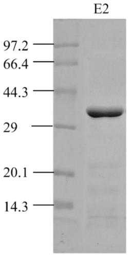
[0038] Referring to the HNJZ-S1 sequence, primers were designed for the E2 protein to amplify the sequence from position 1 to position 304 by PCR (GenBank accessi...
Embodiment 2
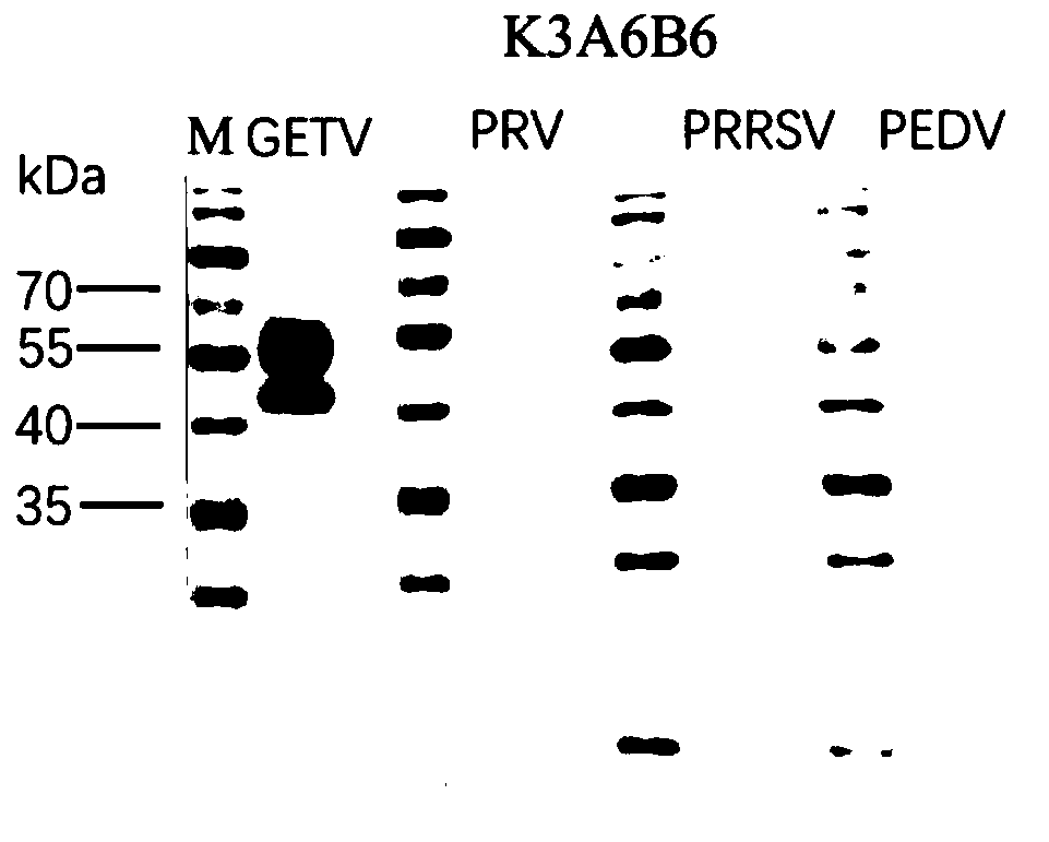
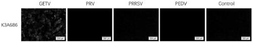
[0097] In this example, the specificity and titer of the monoclonal antibody prepared in Example 1 were tested.
[0098] 1. Western blot experiment
[0099] (1) Get the Marc-145 cells infected by Getavirus, and then lyse them with RIPA lysate. After lysis, centrifuge at 12000g / min for 20min at 4°C; collect the supernatant, and detect the protein concentration of the supernatant with a BCA kit
[0100] (2) Add the supernatant of the cell lysate obtained in step (1) into 12% PAGE, electrophoresis at 120V for 1h; then place the PAGE gel on a PVDF membrane, and transfer to the membrane at 40mA for 90min;
[0101] (3) Put the PVDF membrane of step (2) into 5% skimmed milk, block overnight at 4°C, and then use the K3A6B6 monoclonal antibody of the present invention with a volume ratio of 1:2000 of skimmed milk and monoclonal antibody as the primary antibody , 37°C for 1h;
[0102] (4) Take out the PVDF membrane after step (3), wash it three times with PBST buffer, each time for 3...
Embodiment 3
[0119] 1, E2 protein monoclonal antibody blocking ELISA method
[0120] (1) Purification of Mouse Ascites IgG
[0121] Ascites prepared from the K3A6B6 hybridoma cell line with high titer and specific secretion of anti-Gatavirus monoclonal antibody was taken, purified by Protein G, and the protein content was determined after purification;
[0122] (2) Horseradish peroxidase (HRP) labeled IgG
[0123] The purified ascites IgG was coupled to HRP by simple sodium periodate method;
[0124] (3) Blocking ELISA method
[0125] Take the purified His-E2 fusion protein, dilute it to 5.0 μg / mL (the concentration can also be 2.5 μg / mL, 1.25 μg / mL, etc.) with carbonate buffer solution of pH 9.6, and coat the 96-well ELISA plate , and then incubated at 37°C for 2h;
[0126] After incubation, wash with PBST buffer solution with pH=7.4 for 3 times, then add blocking solution 1% BSA (blocking solution can also be 5% skimmed milk, 1% OVA, 2% gelatin, etc.) After blocking for 1 hour, wash...
PUM
 Abstract
Description
Claims
Application Information