
Method for rapidly detecting retrovirus by using cell ultrathin section electron microscope

A retrovirus and ultra-thin section technology, which is applied in the field of rapid detection and judgment of unknown virus morphology, rapid cell ultra-thin section electron microscope detection of retroviruses, and can solve the problem that the ultra-thin section electron microscope technology cannot be quickly detected and prepared. Complicated, unapplied and other problems, to achieve the effect of fast detection method, reduce interference factors, reduce time cost and labor cost

Pending Publication Date: 2020-08-14
SUZHOU YAOMING KANGDE INSPECTION TESTING
View PDF7 Cites 1 Cited by
  • Summary
  • Abstract
  • Description
  • Claims
  • Application Information

AI Technical Summary

Problems solved by technology

However, due to the complexity of the traditional ultra-thin section sample preparation method, the cycle is long, and a lot of manpower and time costs are required, the rapid detection of ultra-thin section electron microscopy technology cannot be realized, which also makes ultra-thin section electron microscopy technology unable to be used in the highly competitive pharmaceutical industry. better application in the field

Method used


Image

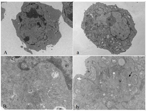
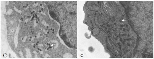
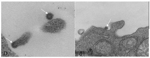
  • Method for rapidly detecting retrovirus by using cell ultrathin section electron microscope
  • Method for rapidly detecting retrovirus by using cell ultrathin section electron microscope
  • Method for rapidly detecting retrovirus by using cell ultrathin section electron microscope

Examples


Embodiment 1

[0036] 1. Experimental steps:

[0037] The present invention provides a kind of fast cell ultrathin section electron microscope detection retrovirus technology, and it comprises the following steps:

[0038] Step 1: Gather 1-2×10 7 Each cell sample was washed 3 times with 1×PBS, fixed with 5% glutaraldehyde for 12 hours, and then fixed with 1% osmic acid for 30 minutes;

[0039] Step 2: Stain with uranyl acetate block and place in a refrigerator at 2-8°C for 2-3 hours to reduce the shaking of the sample;

[0040] Step 3: Acetone dehydration and overtreatment, 70% acetone, 90% acetone, and 100% acetone are dehydrated for 5 minutes each, and should be placed on a shaker during dehydration;

[0041] Step 4: Acetone: Epoxy resin = 1:1 Infiltrate for 20 minutes and then pure resin infiltrate for 2 hours. This step needs to be placed on a shaker;

[0042] Step 5: Polymerize the sample in pure resin at 60°C for at least 10 hours;

[0043] Step 6: Ultra-thin slices of 70-100nm, ca...


PUM


Abstract

The invention discloses a method for rapidly detecting retrovirus by a cell ultrathin section electron microscope. The method comprises the following steps: 1) collecting and fixing of a sample; 2) uranium acetate block dyeing; 3) dehydrating, permeating and polymerizing; 4) slicing, dyeing and detecting through a transmission electron microscope. Compared with a traditional method, the method hasthe advantages that the sample preparation efficiency is greatly improved, the traditional 5-7-day sample preparation period is shortened to 1 day, and an ultrathin slicing technology for rapidly detecting viruses becomes possible. Compared with a traditional virus detection method, the optimized ultrathin section electron microscope technology for detecting viruses has the advantage that the specificity, accuracy and detection efficiency of virus detection are greatly improved.

Description

technical field [0001] The invention relates to the field of biology and medical health, in particular to the field of safety detection of biological products and the rapid detection and judgment of unknown virus morphology, and in particular to a method for rapid cell ultrathin section electron microscope detection of retroviruses. Background technique [0002] Biological products are products made from microorganisms, cells, animal or human tissues and body fluids, etc., using traditional or modern biotechnology and used for the prevention, treatment and diagnosis of human diseases. Human biological products include: bacterial vaccines (including toxoids), viral vaccines, antitoxins and antiserum, blood products, cytokines, growth factors, enzymes, in vivo and in vitro diagnostic products, and other biologically active agents, such as toxins, Antigens, allergens, monoclonal antibodies, antigen-antibody complexes, immune regulation and probiotics, etc. [0003] During the ...

Claims


Application Information

Patent Timeline
Patent Type & Authority Applications(China)
IPC IPC(8): G01N23/04G01N1/28G01N1/30
CPCG01N23/04G01N1/28G01N1/30G01N2001/302G01N2223/102G01N2223/612
Inventor 张宾孙莉王蕾
Owner SUZHOU YAOMING KANGDE INSPECTION TESTING
Learn More