3D printed bio-ink based on costal cartilage as well as preparation method and application thereof

A 3D printing and bio-ink technology, applied in the field of biomedical materials, can solve the problems of waste of donor materials in engraving surgery, large demand for rib bone extraction, and high exposure rate of biocompatibility

Active Publication Date: 2020-09-18
SICHUAN UNIV
View PDF12 Cites 6 Cited by
  • Summary
  • Abstract
  • Description
  • Claims
  • Application Information

AI Technical Summary

Problems solved by technology

[0005] Autologous costal cartilage sculpting surgery is currently the most important surgical method for the treatment of microtia. It has been carried out in major hospitals at home and abroad, but it still has many shortcomings: ① This surgery requires high technical requirements for doctors, and ordinary doctors need to undergo long-term and extensive procedures. Accumulate surgical experience in surgical operations to improve auricle carving techniques; ②The precision of sculpting the outer ear auricle is limited, and there is still a big gap with normal ears; ③The costal cartilage carving surgery requires a large amount of rib bone, and 1-2 costal cartilages are usually Not enough, sculpting surgery often results in wastage of donor material
There have been reports in the literature on the application of synthetic biomaterials such as silica gel and porous polyethylene (Medpor) implanted to reconstruct the auricle of the outer ear. However, due to their biocompatibility and high exposure rate, the application of synthetic biomaterials is limited. limit

Method used


Image

  • 3D printed bio-ink based on costal cartilage as well as preparation method and application thereof

Examples


Embodiment 1

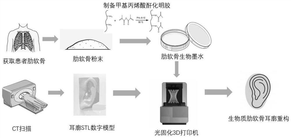
[0052] (1) Costal cartilage biological crushing.

[0053] Take the patient's 7th and 8th costal cartilage (the sixth costal cartilage can be taken if necessary), and use a biological pulverizer to pulverize the costal cartilage in a sterile environment. .

[0054] (2) Configuration of autologous costal cartilage bioink containing photoinitiator

[0055] 20g of gelatin was dissolved in 150mL of alkaline buffered saline solution (0.075mol / L sodium carbonate, 0.175mol / L sodium bicarbonate) at 60°C, then 2mL of methacrylic anhydride was slowly added dropwise to the gelatin solution, and in Stir vigorously at 50 °C for 3 h. After the reaction was completed, 200 mL of deionized water was added to terminate the reaction, and the supernatant was removed by centrifugation, and then the crude product was dialyzed in ultrapure water at 40° C. for 3 days (the cut-off of the dialysis bag was 12-14 KDa). The dialyzed product was then freeze-dried for 3 days to obtain a white porous spong...

Embodiment 2

[0061] In this example, the effects of different autologous costal cartilage bioink formulations on the accuracy of auricle shaping and cartilage remodeling ability were studied. Firstly, the costal cartilage is bio-pulverized, and the 7th and 8th costal cartilages of the patient are taken and crushed with a bio-pulverizer in a sterile environment. The steps and process parameters in this example are the same as those in Example (1), except that the autologous ribs in step (1) have a larger particle size after biological pulverization, and the particle size of the finished product is about 30-80 μm. The effect of its implementation is that the accuracy and resolution of the autologous costal cartilage auricle formed by step (4) 3D printing is worse than that of Example 1, and the order of its forming accuracy is: Example 1>Example 2, this is because the biologically crushed ribs of the printing ink The raw material is rougher, but in this case the pinna has better cartilage re...

Embodiment 3

[0063] In this example, the effects of different autologous costal cartilage bioink formulations on the accuracy of auricle shaping and cartilage remodeling ability were studied. Firstly, the costal cartilage is bio-pulverized, and the 7th and 8th costal cartilages of the patient are taken, and the costal cartilage is crushed with a bio-pulverizer in a sterile environment. The steps and process parameters in this example are the same as those in Example (1), except that the autologous ribs in step (1) have a larger particle size after bio-crushing, and the particle size of the finished product is about 80-130 μm. The effect of its implementation is that the accuracy and resolution of the autologous costal cartilage auricle formed by 3D printing in step (4) is worse than that of Example 2, and the order of its forming accuracy is: Example 1>Example 2>Example 3, this is because the printing ink The bio-crushed rib raw material is rougher, but the auricle of this example has bett...


PUM

PropertyMeasurementUnit
particle sizeaaaaaaaaaa

Abstract

The invention discloses 3D printed bio-ink based on costal cartilage as well as a preparation method and application thereof, and belongs to the technical field of biomedical materials. The 3D printedbio-ink comprises costal cartilage powder, photocurable hydrogel and phenyl-2,4,6-trimethylbenzoyl lithium phosphonate, and an artificial auricle can be successfully prepared by adopting the 3D printed bio-ink through a reverse engineering system and a photocurable 3D printing technology. The 3D printed autologous costal cartilage auricle prepared by the 3D printed bio-ink has the advantages of high adaptive individuation, simple and fast preparation process, high appearance precision, full utilization of donor autologous materials, great reduction of costal cartilage taking amount and good biocompatibility and tissue reconstruction capability, and has great clinical application value in a microtia reconstruction operation.

Description

technical field [0001] The invention belongs to the technical field of biomedical materials, in particular to a reverse engineering method from CT scanning image data to auricle production, and in particular to a preparation method of autologous costal cartilage bio-ink and its molding and manufacturing technology. [0002] technical background [0003] Congenital microtia (Congenital microtia) is a type of congenital disease characterized by severe hypoplasia of the pinna, atresia or stenosis of the external auditory canal, and deformity of the middle ear. According to statistics, the incidence of microtia is about 1 / 3000-1 / 5000 in the oriental population. Its clinical symptoms and accompanying deformities not only cause the appearance and hearing of the children and other organ dysfunction, but also cause the children and their families to have serious problems. a great psychological burden. Congenital microtia is usually treated by auricle reconstruction. Due to the compl...

Claims


Application Information

Patent Timeline
Patent Type & Authority Applications(China)
IPC IPC(8): A61L27/36A61L27/20A61L27/22A61L27/52A61L27/58B33Y10/00B33Y70/10
CPCA61L27/20A61L27/222A61L27/227A61L27/3612A61L27/3654A61L27/52A61L27/58A61L2430/14B33Y10/00B33Y70/10C08L5/08C08L89/00
Inventor 李正勇周长春唐佩宋平吴丽娜彭智愚雷皓远范洪远蒋青樊渝江张兴栋
Owner SICHUAN UNIV
Why Eureka
Try Eureka