Method for real-time spatial positioning of ablation catheter in heart cavity by using ultrasound

An ablation catheter and spatial positioning technology, which is applied in the field of medical devices, can solve the problems of insufficient angle adjustment, cumbersome process, and many instruments and equipment, and achieves the effect of facilitating adjustment and improving detection efficiency.

Inactive Publication Date: 2020-10-02
JIANGSU TINGSN TECH CO LTD
View PDF0 Cites 5 Cited by
  • Summary
  • Abstract
  • Description
  • Claims
  • Application Information

AI Technical Summary

Problems solved by technology

Since the existing technology realizes the real-time visualization of the spatial position of the ablation catheter through electric / magnetic field positioning, and then realizes the three-dimensional modeling of the heart through point-to-point software algorithms, and then obtains the positioning of the ablation catheter in the heart, although this method can be real-time and accurate The location of the ablation catheter in the heart, but the whole process requires many instruments and equipment, and the process is cumbersome
[0003] Moreover, the current device needs multi-angle adjustments when in use, and the current device often encounters the situation that the angle adjustment is not in place when the current device is used, resulting in inaccurate detection angles, making it difficult to accurately detect patients, which is not conducive to medical analysis and develop medical plans

Method used


Image

  • Method for real-time spatial positioning of ablation catheter in heart cavity by using ultrasound
  • Method for real-time spatial positioning of ablation catheter in heart cavity by using ultrasound
  • Method for real-time spatial positioning of ablation catheter in heart cavity by using ultrasound

Examples


Embodiment 1

[0030] see figure 2 A flow chart of a method for real-time spatial positioning of an ablation catheter in a cardiac cavity provided by the present application; including the following steps:

[0031] Obtain an ultrasound device to scan the sectors of the heart at different angles using two-dimensional ultrasound to reconstruct a three-dimensional model of the heart;

[0032] In the three-dimensional model, the real-time three-dimensional space positioning of the ablation catheter in the heart is realized.

Embodiment 2

[0034] Obtain the ultrasonic device installed on the electrode in the coronary sinus and use two-dimensional ultrasound to scan the sectors of the heart at different angles to reconstruct the three-dimensional model of the heart;

[0035] In the three-dimensional model, the real-time three-dimensional space positioning of the ablation catheter in the heart is realized.

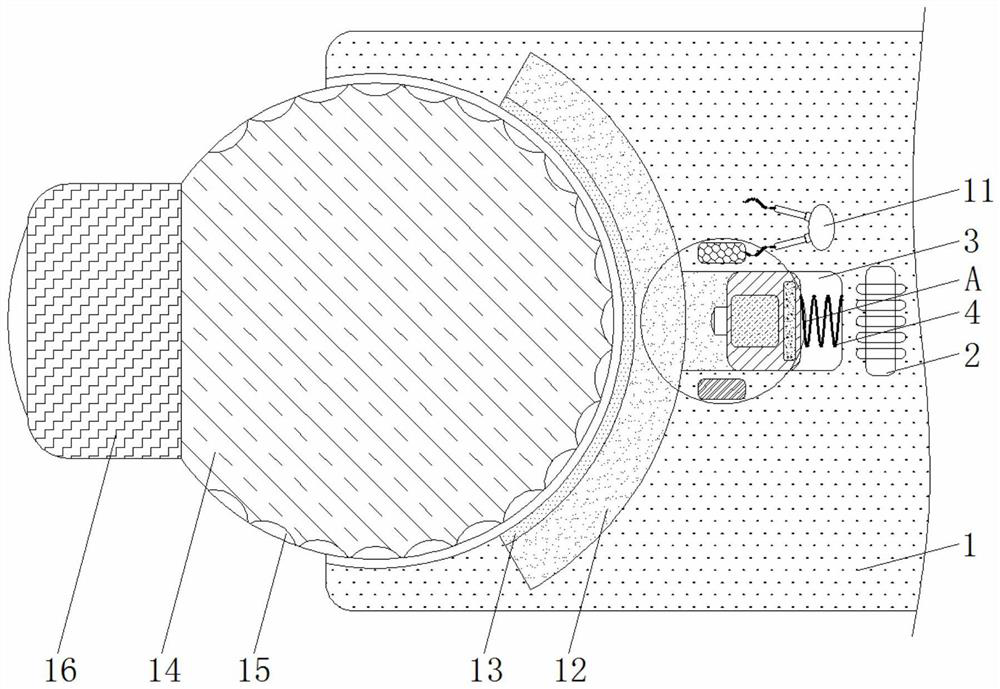
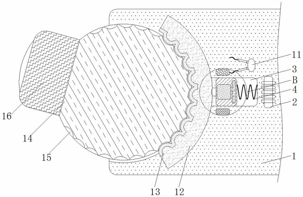
[0036] The coronary sinus is a C-shaped vein that circles the surface of the heart (a heart wall about 10mm thick from the heart cavity), opening in the right atrium, with an inner diameter of about 3-5mm and a length of 10-12mm, such as image 3 as shown, Figure 8 The placement position of the ultrasonic device provided in Example 1 of the present invention in the coronary sinus.

[0037] During the operation, a motor needs to be inserted into the coronary sinus for diagnosis, and an ultrasound device is installed on the electrodes in the coronary sinus to realize real-time three-dimensional spatial positioni...

Embodiment 3

[0039] Obtain an ultrasound device set on the esophagus adjacent to the heart and use two-dimensional ultrasound to scan the sectors of the heart at different angles to reconstruct a three-dimensional model of the heart;

[0040] In the three-dimensional model, the real-time three-dimensional space positioning of the ablation catheter in the heart is realized.

[0041] see Figure 9 , is the placement position of the ultrasonic device provided in this application in the esophagus. The esophagus is anatomically attached to the posterior wall of the heart, as shown in the figure circled by the line. Therefore, the ultrasonic device is installed on the esophagus to realize real-time three-dimensional spatial positioning of the ablation catheter in the heart.


PUM


Abstract

The invention discloses a method for real-time spatial positioning of an ablation catheter in a heart cavity by using ultrasound, and relates to the technical field of medical instruments. A fixing seat is included, the right inner wall of the fixing seat is fixedly connected with an electromagnet, and a buffer groove is formed in the right inner wall of the fixing seat and close to the left sideof the electromagnet. According to the method for real-time spatial positioning of the ablation catheter in the heart cavity by using ultrasound, through cooperative use of the electromagnet and a resistor, during use of the device, the electromagnet is energized first, so that a piston block moves leftwards under the mutual repulsive force of the electromagnet and a magnet to squeeze an electrorheological fluid to the left side, a rubber ring expands and is clamped with a clamping groove, when the circuit voltage is larger than the minimum path voltage of the resistor, the electrorheologicalfluid becomes solid at the moment to fix a universal ball, so that fixation of an ultrasound device is effectively guaranteed, when the electrorheological fluid is liquid, the universal ball can be adjusted by universal rotation, and the effects of facilitating the adjustment of the angle of the ultrasound device and improving the detection efficiency are achieved.

Description

technical field [0001] The invention relates to the technical field of medical devices, in particular to a method for real-time spatial positioning of an ablation catheter in a cardiac cavity by using ultrasound. Background technique [0002] The three-dimensional platform has become the mainstream of modern electrophysiology and radiofrequency ablation surgery. The three-dimensional mapping systems currently used in clinical practice are all based on magnetic field positioning, electric field positioning, and magnetic field and electric field dual positioning. The three electric / magnetic fields emit three different fields, and the real-time three-dimensional spatial positioning is realized by calculating the relative position changes of the ablation catheter tip in the three fields in real time. Specifically, the field emission device realizes the real-time visualization of the spatial position of the ablation catheter through electric / magnetic field positioning, and then r...

Claims


Application Information

Patent Timeline
Patent Type & Authority Applications(China)
IPC IPC(8): A61B34/20A61B34/10A61B18/00
CPCA61B2018/00351A61B2018/00577A61B34/10A61B34/20A61B2034/102A61B2034/107A61B2034/2063
Inventor 赵东生王静吴建张冬宇
Owner JIANGSU TINGSN TECH CO LTD
Why Eureka
Try Eureka