Application method of nafamostat mesylate to treatment of chlamydia infection of genital tract

A technology for nafamostat mesylate and reproductive tract infection, applied in the field of biomedicine

Inactive Publication Date: 2020-10-27
THE SECOND XIANGYA HOSPITAL OF CENT SOUTH UNIV
View PDF3 Cites 0 Cited by
  • Summary
  • Abstract
  • Description
  • Claims
  • Application Information

AI Technical Summary

Problems solved by technology

[0005] At present, there is no report on the application of nafamostat mesylate in the treatment of chlamydia reproductive tract infection and the prevention of complications such as tubal obstruction, hydrosalpinx, and tubal infertility

Method used


Image

  • Application method of nafamostat mesylate to treatment of chlamydia infection of genital tract
  • Application method of nafamostat mesylate to treatment of chlamydia infection of genital tract
  • Application method of nafamostat mesylate to treatment of chlamydia infection of genital tract

Examples


Embodiment 1

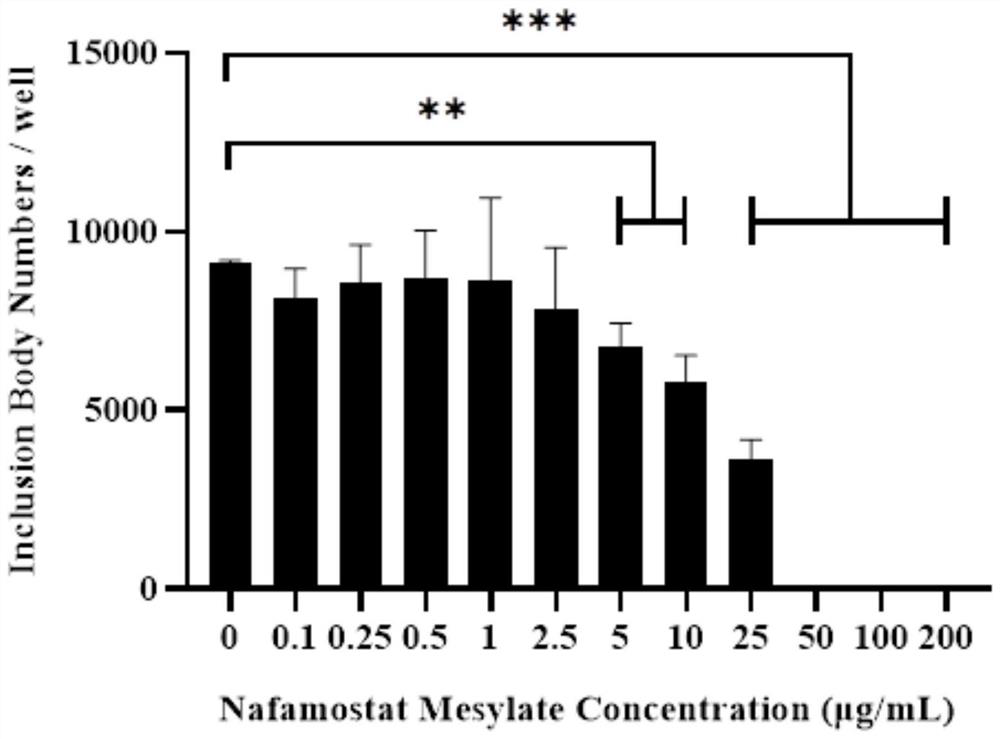
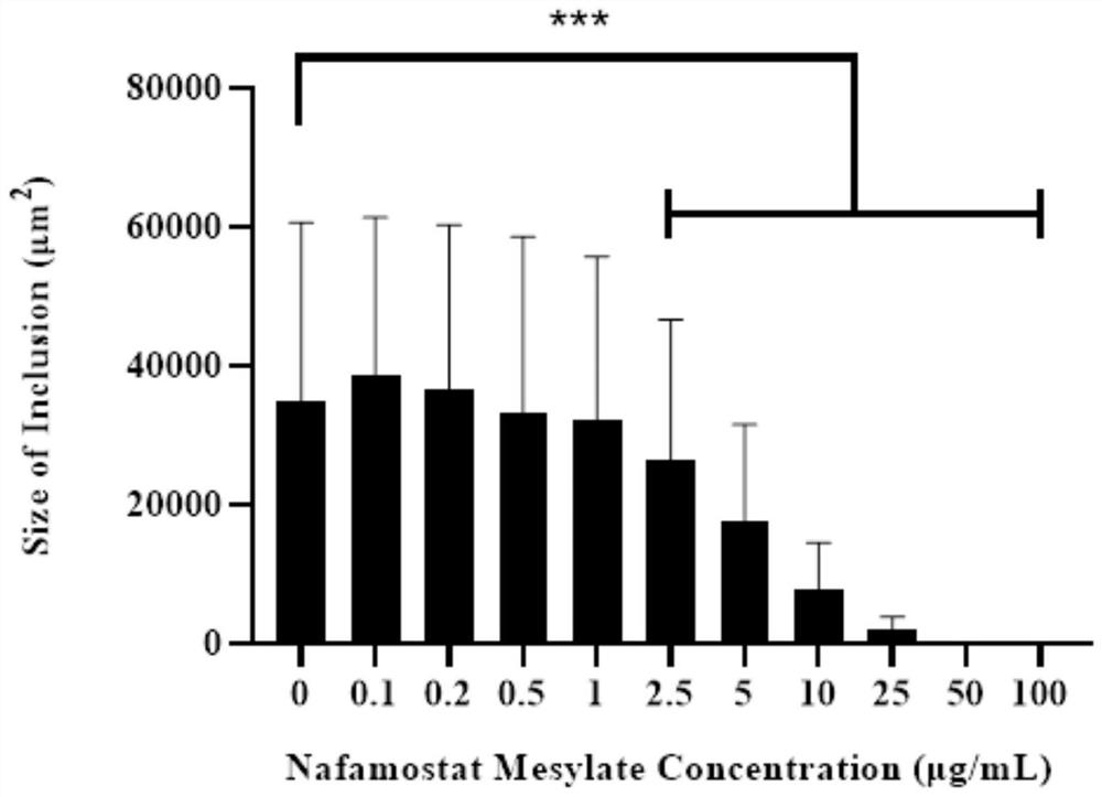
[0045] Inhibitory effect of nafamostat mesylate on genital tract epithelial cells infected by chlamydia

[0046] 1. Experimental materials

[0047] Strains and cells: Chlamydia muridarum (Cm) Cm-mCherry fluorescent strain, constructed by our laboratory, routinely passaged, purified, and preserved. The fluorescent strain introduced the red fluorescent protein mCherry gene into the plasmid of wild-type Cm. The strain comes with Fluorescence, without fixation and antibody incubation, can be used for fluorescence observation in vivo after infection of cells. HeLa229 cells were purchased from the American Culture Collection (ATCC), and were routinely passaged and preserved by our laboratory;

[0048] Main reagents: nafamostat mesylate was purchased from Abcam Company of the United States; high-glucose DMEM medium and trypsin were purchased from Hyclone Company of the United States; fetal bovine serum was purchased from Gibco Company of the United States; cycloheximide, gentamicin,...

Embodiment 2

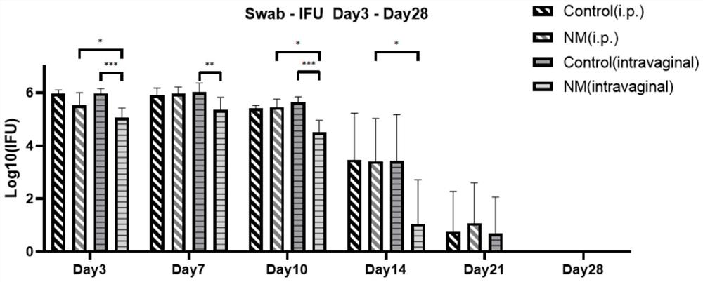
[0056] Effects of Nafamostat Mesylate on the Reproductive Tract of Mice Infected with Chlamydia

[0057] 1. Experimental materials

[0058] Animal: C57BL / 6 mouse, SPF grade, 6 weeks old, female, purchased from Hunan Slake Jingda Experimental Animal Co., Ltd., license number: SCXK (Xiang) 2019-0004;

[0059] Cells and main reagents are the same as in Example 1.

[0060] 2. Experimental method

[0061] Grouping of animals: 5 animals in the abdominal control group, 5 animals in the vaginal control group, 6 animals in the intraperitoneal administration group, and 7 animals in the vaginal administration group.

[0062] Infection model establishment: 5 days before infection with Chlamydia (-5d), each mouse was pre-injected with 2.5 mg of medroxyprogesterone subcutaneously to synchronize the estrous cycle of the mice and facilitate Chlamydia infection. On the day of infection (0d), each mouse was inoculated vaginally with 5×10 5 Chlamydia inclusion body forming units (Inclusion f...

Embodiment 3

[0070] Effects of intravaginal infusion of nafamostat mesylate on cytokines in the upper and lower reproductive tracts of mice infected with chlamydia

[0071] 1. Experimental materials

[0072] Animals, cells, and main reagents are the same as in Example 2.

[0073] 2. Experimental method

[0074] Grouping of animals: 10 vaginal control groups and 10 vaginal administration groups;

[0075] The establishment of the infection model and the way of administration are the same as in Example 2.

[0076] Determination of cytokines in the upper and lower reproductive tracts: 5 mice in each group were randomly killed on the 7th and 14th day after infection, and the upper and lower reproductive tracts were carefully separated. Add upper and lower reproductive tract tissues and 500 μl SPG buffer respectively to the homogenizer, grind on ice, collect the homogenate, centrifuge at 4° C., 500 g for 5 minutes, and collect the supernatant. Use BD TM Cytometric Bead Array kit (BD CBA mo...


PUM

PropertyMeasurementUnit
Concentrationaaaaaaaaaa

Abstract

The invention relates to the technical field of biomedicine, in particular to an application method of nafamostat mesylate to treatment of chlamydia infection of a genital tract. The application method comprises the following steps of: A, selecting experimental materials; B, developing an experimental method; C, observing experimental results; and D, drawing experimental conclusions. The inventionprovides a new application of the nafamostat mesylate to treatment of chlamydia infection of the genital tract and particularly relates to an application of the nafamostat mesylate to preparation ofdrugs for treating chlamydia infection of the genital tract, and an application of the nafamostat mesylate to preparation of drugs for preventing tubal obstructions, hydrosalpinx and tubal infertilitycaused by chlamydia infection of the genital tract. Through in vitro cell experiments, animal experiments and other aspects, it is proved that the nafamostat mesylate can effectively treat chlamydiainfection of the genital tract to prevent and treat a tubal infertility disease.

Description

technical field [0001] The invention relates to the technical field of biomedicine, in particular to an application method of nafamostat mesylate in treating chlamydia reproductive tract infection. Background technique [0002] Chlamydia (Chlamydia) is a group of extremely small, non-motile, microorganisms that grow exclusively in cells. The genitourinary tract infection caused by it is the most common sexually transmitted disease worldwide. According to WHO statistics, there are new Approximately 127.2 million additional patients [1] . Because the clinical manifestations of chlamydia infection are non-specific, especially in female infected patients, asymptomatic patients account for about 80% of the total, so it is often missed in clinical diagnosis. If patients are not diagnosed and treated in time, they are prone to develop chronic and persistent infection, causing damage, adhesion and scar formation of reproductive tract tissue, and then causing many complications suc...

Claims


Application Information

Patent Timeline
IPC IPC(8): A61K31/245A61P31/04A61P15/08A61P15/02
CPCA61K9/0034A61K31/245A61P15/02A61P15/08A61P31/04
Inventor 陈建林彭亮张宏波沈宏伟唐玲丽胡紫昊赵宇婕刘珊珊鲁琼
Owner THE SECOND XIANGYA HOSPITAL OF CENT SOUTH UNIV
Why Eureka
Try Eureka