Method for constructing phage display multivariate cyclic peptide library based on disulfide bond precise pairing

A phage display and disulfide bond technology, which is applied in the field of constructing phage display multi-cyclic peptide libraries based on disulfide bond precise pairing, can solve the problems of poor tolerance to sequence modification and complex oxidation folding process, and achieve high selectivity and affinity , high oral availability and strong metabolic stability

Active Publication Date: 2020-11-17
XIAMEN UNIV
View PDF4 Cites 2 Cited by
  • Summary
  • Abstract
  • Description
  • Claims
  • Application Information

AI Technical Summary

Problems solved by technology

[0005] The purpose of the present invention is to provide a method for constructing a phage display multi-cyclic peptide library based on precise pairing of disulfide bonds in view of the defects of existing natural disulfide-bond-rich cyclic peptide molecular skeletons, such as complicated oxidative folding process and poor tolerance to sequence modification.

Method used


Image

  • Method for constructing phage display multivariate cyclic peptide library based on disulfide bond precise pairing
  • Method for constructing phage display multivariate cyclic peptide library based on disulfide bond precise pairing
  • Method for constructing phage display multivariate cyclic peptide library based on disulfide bond precise pairing

Examples


Embodiment 1

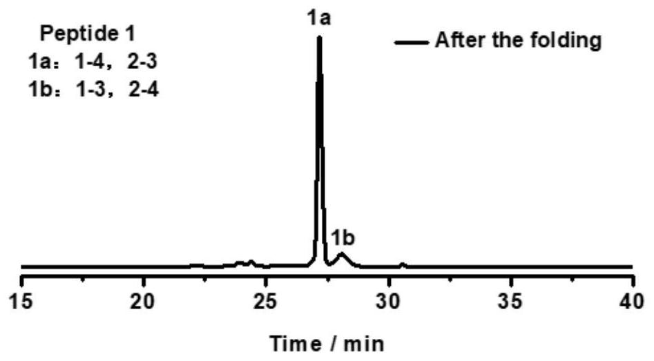
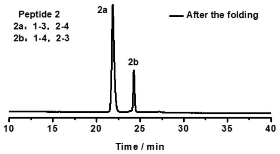
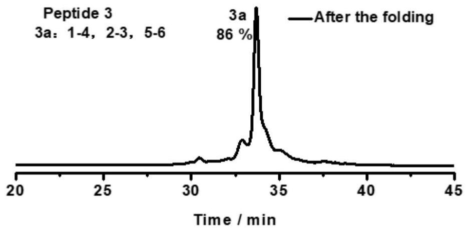
[0093] Take the oxidative folding of peptide 1 as an example (peptide 2, peptide 3, peptide 4, peptide 5, peptide 6 oxidative folding steps are the same as peptide 1, and their chromatograms are as follows Figure 2-6 ), its specific oxidation method is:

[0094] The solid powder of peptide 1 was dissolved in an aqueous solution (containing 0.1% trifluoroacetic acid to prevent oxidation of the peptide) for peptide quantification. The 50 uM polypeptide was placed in a phosphate buffer solution (100 mM pH=7.4) containing 0.5 mM GSSG, and reacted in a shaker at 37° C. for 4 h. The chromatogram of the oxidation product is as figure 1 As shown, two expected products (named 1a and 1b, respectively) were obtained.

Embodiment 2

[0096] In order to determine the pairing mode of oxidation products, taking peptide 1 as an example, a schematic diagram of the determination process is shown ( Figure 7 ). Replace the two cysteines (positions 2 and 3) in peptide 1 with Acm-protected cysteine ​​peptide 7 (sequence: H-WGCPPC(Acm)GGKGGC(Acm)PPCGW-NH 2 ), synthesized and quantitatively placed in a phosphate buffer solution (100 mM pH=7.4) containing 0.5 mM GSSG for oxidation reaction. Utilize high-performance liquid chromatography to purify and separate the product after lyophilization, dissolve in methanol (containing 1% trifluoroacetic acid), then add twice equivalent or ten times equivalent of iodine to carry out de-Acm protection and form disulfide bond at the same time, the obtained oxidation The product is the pairing method of (1-4,2-3). The chromatographic position of the product is determined by high performance liquid chromatography, and the chromatographic position is compared with the oxidation prod...

Embodiment 3

[0098] The schematic diagram of the construction of phage display three-membered cyclic peptide library is as follows: Figure 8 shown. First, the chemically synthesized library DNA fragments and extension primers were reacted in a metal bath at 95°C for 3 minutes, and then quickly cooled on ice for 5 minutes. Afterwards, Klenow enzyme was added to the above-mentioned return system to extend the reaction for 4 hours in a 37°C metal bath, and then placed in a 65°C metal bath to inactivate the enzyme activity. Then the extension product and phagemid vector pCantab 5E were digested with SfiI (12h, 50°C) and NotI (12h, 37°C). The extension products and phagemid vectors after SfiI / NotI double digestion were purified by polyacrylamide gel electrophoresis (PAGE) and agarose gel electrophoresis, respectively. After purification, the extension products and phagemid vectors were purified at a ratio of 1:10 Molar Ratio The ligation reaction was carried out overnight (16h) at a temperat...


PUM


Abstract

The invention discloses a method for constructing a phage display multi-element cyclic peptide library based on disulfide bond precise pairing, and relates to cyclic peptide compounds. A disulfide bond pairing mode in a ternary cyclic peptide molecule is accurately regulated and controlled, a ternary cyclic peptide template is fused and expressed on the surface of phage, and a phage ternary cyclicpeptide library is constructed; besides, two CPPC motifs and two cysteines are inserted into a polypeptide skeleton by utilizing the tendency property that the CPPC motifs have preferential generation of parallel paired dimers, so that pairing of disulfide bonds and polypeptide folding are effectively regulated and controlled, and by changing the arrangement positions of the two CPPC motifs and the two cysteines, the disulfide bonds can be effectively regulated and controlled. Therefore, the novel disulfide bond-rich cyclic peptide compounds with different polycyclic topological structures can be designed from origin; and the phage three-membered cyclopeptide ligand can be used as the template of the polycyclic peptide library without the insertion of the natural cyclopeptide skeleton andthe non-natural amino acid; therefore, the phage three-membered cyclopeptide library is successfully constructed by using the template, and the functional cyclopeptide ligand with the new structure,which is not derived from the natural product scaffold structure, is screened out.

Description

technical field [0001] The present invention relates to cyclic peptide compounds, in particular to a method for constructing a phage display multi-cyclic peptide library based on precise pairing of disulfide bonds. Background technique [0002] Cyclic peptide compounds are a class of cyclic compounds with special structure, stable conformation, wide range of biological activities, and unique mechanism of action. They have high binding affinity and specificity for target receptors, low cytotoxicity, and high oral availability. Therefore, the research and development of ligand drugs for cyclic peptide compounds has attracted more and more attention. For example, the cyclic peptide drugs Telavancin and Dalbavancin for bacterial and fungal infections; the anti-tumor cyclic peptide drugs Lanreotide and Romidepsin, these drugs have been successfully marketed and have exerted great medical value. However, among these drugs, especially some cyclic peptide drugs rich in disulfide bo...

Claims


Application Information

Patent Timeline
Patent Type & Authority Applications(China)
IPC IPC(8): C40B50/06C12N15/70C40B40/02C12R1/19
CPCC40B50/06C12N15/70C40B40/02
Inventor 吴川六路帅敏吴亚培
Owner XIAMEN UNIV
Why Eureka
Try Eureka