Double-fluorescent-protein positioning detection system for detecting mitochondrial autophagy of cells and application

A technology of mitophagy and dual fluorescent protein, applied in the biological field, can solve the problems of cumbersome process, cumbersome electron microscope detection process, long cycle, etc., and achieve the effect of solving the cumbersome operation process

Pending Publication Date: 2021-01-01
SHANGHAI BIOCHIP
View PDF0 Cites 2 Cited by
  • Summary
  • Abstract
  • Description
  • Claims
  • Application Information

AI Technical Summary

Problems solved by technology

[0004] However, the research method of mitophagy is still not perfect. The most intuitive detection method of mitophagy at present is to observe the specific autophagosome morphology through electron microscopy or to observe the key protein Lc3b (microtubule associated protein 1light) on the autophagosome by immunofluorescence experiment. chain 3beta) is located on the mitochondria, but the electron microscopy detection process is cumbersome and the cycle is long, the observation area is limited, the antibody titer of immunofluorescence is high, the experiment cycle is long, and the process is cumbersome
Mitochondrial dyes currently on the market are only effective for staining living cells, and are not ideal for experiments involving cell fixation and permeabilization, such as immunofluorescence.
Moreover, the fluorescence of antibodies and dyes is easily quenched, and the shooting effect is not good

Method used


Image

  • Double-fluorescent-protein positioning detection system for detecting mitochondrial autophagy of cells and application
  • Double-fluorescent-protein positioning detection system for detecting mitochondrial autophagy of cells and application
  • Double-fluorescent-protein positioning detection system for detecting mitochondrial autophagy of cells and application

Examples


Embodiment 1

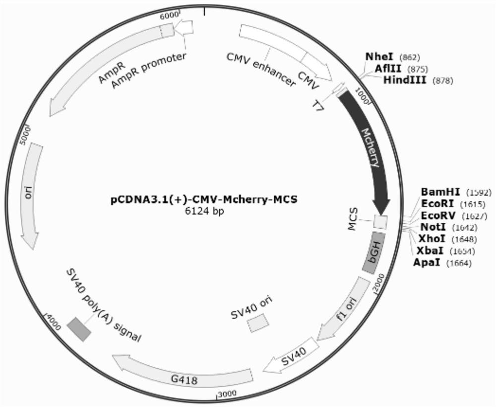
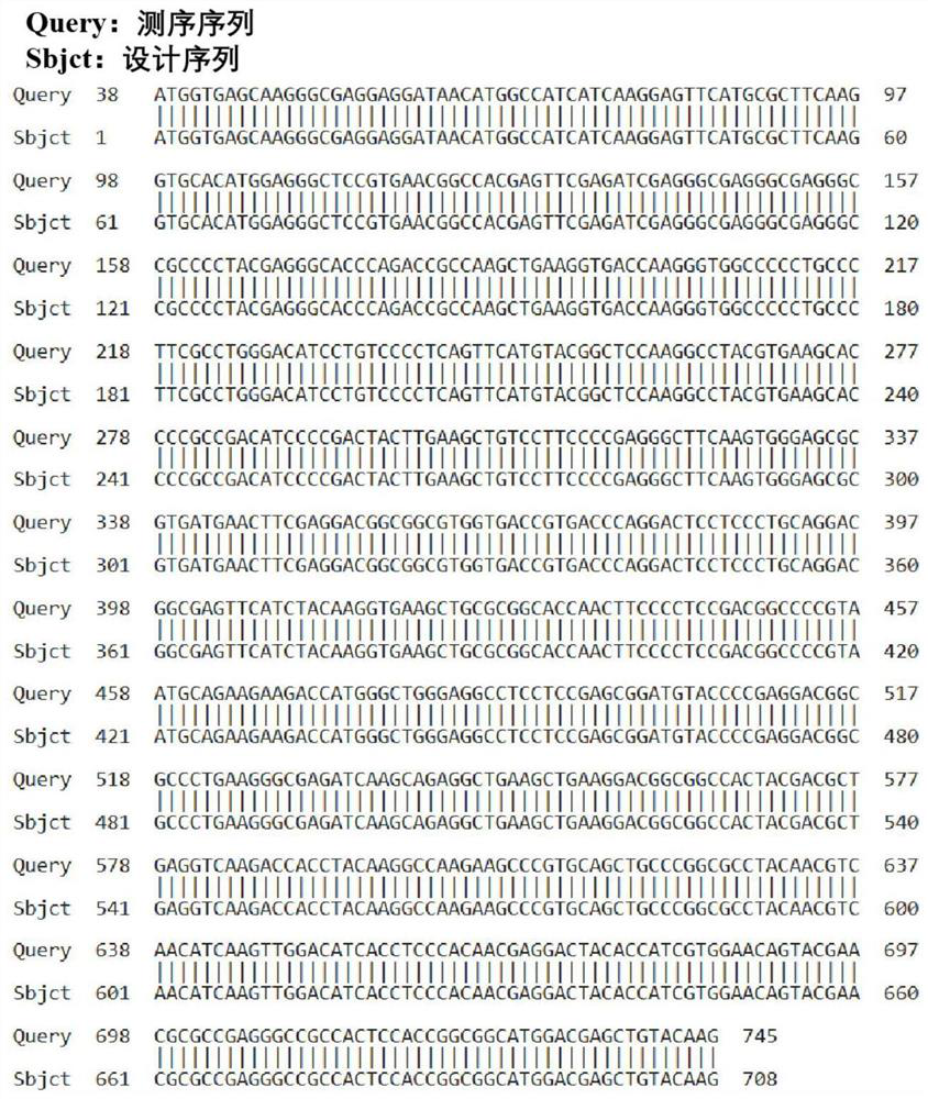
[0077] The pcDNA3.1(+) empty plasmid was used to design Mcherry primers, the PCR products were tapped and recovered, and the pcDNA3.1(+)-Mcherry-MCS fusion expression plasmid was constructed, identified by enzyme digestion, sequence comparison, and expression identification.

[0078] Using the sequence of the PcDNA3.1-Tom20-Mcherry plasmid as a template (remove the TAA stop codon), select the HindⅢ and BamhI restriction sites on the pcDNA3.1(+) empty plasmid, and use the Primer 5 software to design Mcherry primers , and add the corresponding restriction site sequence (HindⅢ: AAGCTT , Bamh I: GGATCC ) and the corresponding protected base sequence (HindⅢ: CCC, BamhⅠ: CG), as follows:

[0079] Mcherry-HindⅢ-F: CCC AAGCTT ATGGTGAGCAAGGGCGAGG; (SEQ ID NO: 3)

[0080] Mcherry-BamhⅠ-R: CG GGATCC CTTGTACAGCTCGTCCATGCC; (SEQ ID NO: 4)

[0081]The target fragment (725 bp) was recovered by 1% agarose gel electrophoresis after PCR reaction of 50 μL system, and the target fragment...

Embodiment 2

[0086] Using the pcDNA3.1(+)-Mcherry-MCS fusion expression plasmid and designing the full-length primer of LC3B, using the cDNA of human lung adenocarcinoma cell line A549 as a template, the PCR product was recovered by tapping rubber to construct pcDNA3.1(+)-Mcherry- Lc3b fusion expression plasmid, enzyme digestion identification, sequencing comparison verification, expression identification.

[0087] Referring to the sequence information of the CDS coding region of Homo sapiens microtubule associated protein 1light chain 3beta (NCBI Reference Sequence: NM_022818.5), using the cDNA of human lung adenocarcinoma cell A549 as a template, select pcDNA3.1(+)-Mcherry-MCS fusion expression EcorI and XbaI restriction sites on the plasmid, use Primer 5 software to design MAP1LC3B primers, and ensure that LC3B will not undergo frameshift mutations (base T is added to the 5' end of the forward primer), and in the forward and reverse Add the corresponding restriction site sequence (EcorI...

Embodiment 3

[0095] Using PcNA3.1-MCS-EGFP fusion to express the empty plasmid, design the full-length primer of PHB1, use the cDNA of human lung adenocarcinoma cell line A549 as a template, and recover the PCR product by rubber tapping to construct the fusion of pcDNA3.1(+)-PHB1-EGFP Expression plasmids, enzyme digestion identification, sequencing comparison, expression identification.

[0096]Referring to Homo sapiens prohibitin (PHB), transcript variant 2 (NCBI ReferenceSequence: NM_002634.4) CDS coding region sequence information (remove stop codon TGA), using the cDNA of human lung adenocarcinoma cell A549 as a template, select pcDNA3.1 ( +)-MCS-EGFP fusion expresses the HindⅢ and EcorⅠ restriction sites on the empty plasmid, and uses Primer 5 software to design MAP1LC3B primers, and ensures that EGFP will not undergo frameshift mutations (add Base C), and add the corresponding restriction site sequence (HindⅢ: AAGCTT, EcorI: GAATTC) and the corresponding protection base sequence (Hin...


PUM


Abstract

The invention discloses a double-fluorescent-protein positioning detection system for detecting mitochondrial autophagy of cells. The detection system at least comprises (1) fusion protein composed offluorescent protein A and indicating protein A; and (2) fusion protein composed of a fluorescent protein B and an indicator protein B, wherein the fluorescent protein A and the fluorescent protein Bhave different fluorescence colors, the indicating protein A is used for indicating autophagosome, and the indicating protein B is used for indicating mitochondria. The invention provides a quicker, more convenient and more accurate way for researching the observation of mitochondrial autophagy. The system is a good supplement for methodology of mitochondrial autophagy research, and opens up a newidea for methodology of mitochondrial autophagy research.

Description

technical field [0001] The invention belongs to the field of biotechnology, and in particular relates to a dual fluorescent protein localization detection system and application for detecting cell mitochondrial autophagy. Background technique [0002] Mitochondria, as the "energy factory" of cell metabolism, is an important place for cellular tricarboxylic acid cycle and oxidative phosphorylation. Whether the mitochondrial function is normal or not is directly related to the damage of cells, tissues and even the body. Therefore, the damaged mitochondria must be effectively removed to ensure the normal life activities of cells. Mitophagy is a dynamic physiological balance process in which cells selectively remove damaged mitochondria to regulate the number of intracellular mitochondria and maintain the normal function of mitochondria. [0003] In recent years, mitophagy has become one of the newly developed research directions of autophagy. A large number of research repor...

Claims


Application Information

Patent Timeline
Patent Type & Authority Applications(China)
IPC IPC(8): G01N33/68G01N33/533C12N15/62
CPCC07K14/43595C07K2319/00G01N33/533G01N33/68
Inventor 苏军王珂徐祎春周佳菁赵英楠马晓卯冯晨晨
Owner SHANGHAI BIOCHIP
Why Eureka
Try Eureka