Eureka AIR delivers breakthrough ideas for toughest innovation challenges, trusted by R&D personnel around the world.

Method and device for computed tomography imaging

A tomography, computerized technology used in computed tomography scanners, instruments for radiological diagnosis, computer-aided medical procedures, etc., to solve problems such as high bleeding risk and inability to reliably predict bleeding in the infarcted area of ​​a patient

Pending Publication Date: 2021-01-19
SIEMENS HEALTHCARE GMBH
View PDF0 Cites 1 Cited by
  • Summary
  • Abstract
  • Description
  • Claims
  • Application Information

AI Technical Summary

Problems solved by technology

Therefore, it is not possible to reliably predict whether a patient will experience infarct-area hemorrhage if thrombolytic therapy is administered after a time window beyond 4.5 hours
Previous studies have shown that there is a higher risk of bleeding in this condition

Method used


Image

  • Method and device for computed tomography imaging
  • Method and device for computed tomography imaging
  • Method and device for computed tomography imaging

Examples


Embodiment Construction

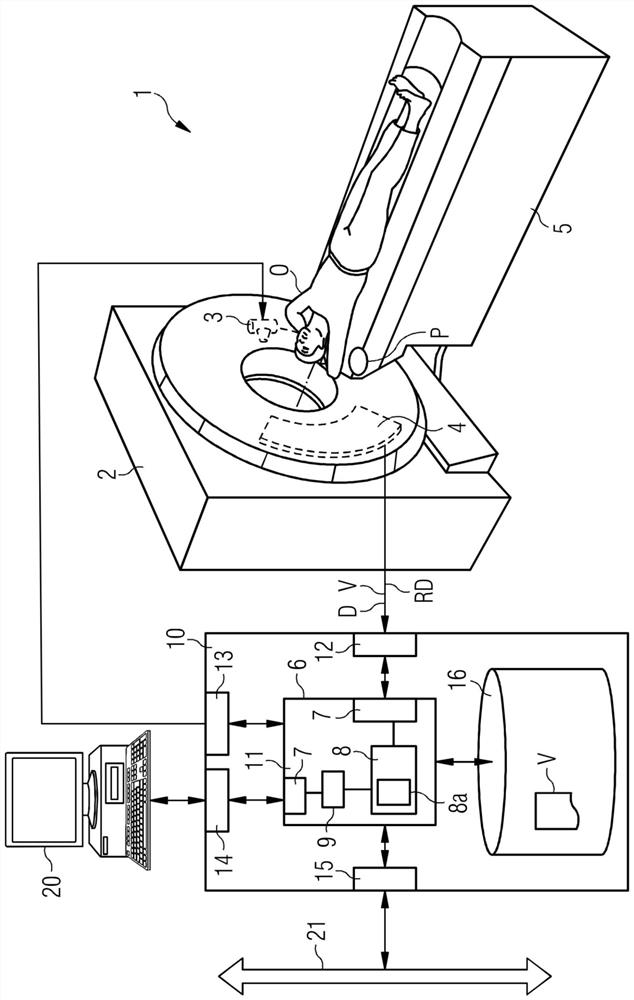
[0094] figure 1A computed tomography system 1 is shown schematically with a control device 10 for carrying out the method according to the invention. Here, the CT system 1 is configured as a photon counting system and has, in a conventional manner, a scanner 2 with a gantry in which an x-ray source 3 rotates, which respectively transmits a patient, who The couch 5 is pushed into the measurement space of the gantry, so that the radiation impinges on a detector 4 , which is a photon-counting detector, located opposite the x-ray source 3 . It should be clearly pointed out that, according to the figure 1 The embodiment of is just one example of CT, and the invention can also be used in any multi-spectral recording CT configuration, such as a CT configuration with a ring-shaped fixed X-ray detector and / or multiple X-ray sources.

[0095] For the control device 10 , likewise only those components are shown which are necessary for explaining the invention. In principle, such CT sy...


PUM


Abstract

The invention relates to a method for computed tomography imaging. The method includes provisioning a CT data set (D) of an object (O), the CT data set being previously recorded via a multispectral recording method; suppressing a contrast, caused by a tissue type (G1, G2), and generating a contrast-suppressed data set (D) from the CT data set provisioned; and analyzing at least the contrast-suppressed data set(K) generated or a data set(D) generated via a machine learning algorithm based on the contrast-suppressed data set (K), the analyzing being configured to identify at least one change inthe tissue type (G1, G2). A corresponding device, a control device (10) for a computed tomography system or a diagnosis system, and a diagnosis system and a computed tomography system (1) are also disclosed.

Description

technical field [0001] The present invention relates to a method and apparatus for imaging in computed tomography, and in particular to a method and apparatus for identifying and analyzing different tissue components by utilizing multispectral (photon counting) data recording in combination with iterative data reconstruction and artificial intelligence. Methods for the quantitative assessment of components, especially in the case of cerebral ischemia, to predict the benefit of various treatments. Here, the invention is used for imaging, which means that the invention can be directly applied in the field of image reconstruction, the invention can be used to process the reconstructed image, or can generate in addition to the original data set in the preparatory stage of reconstruction Additional data or additional data about the original dataset. Background technique [0002] To date, it has not been possible to satisfactorily represent all areas of the human body with an app...

Claims


Application Information

Patent Timeline
Patent Type & Authority Applications(China)
IPC IPC(8): A61B6/03G06T11/00G06T17/00G06N3/04G06N3/08
CPCA61B6/032A61B6/5211A61B6/5205A61B6/5217G06T11/003G06T17/00G06N3/08G06N3/045A61B6/482A61B6/583G16H50/20G16H30/40G16H30/20G06T7/0012G06T2207/10081G06T2207/20081G06T2207/20084G06T2207/30016G06T11/008G06T2211/408G06N7/01G06T2211/441A61B6/5258A61B6/501A61B6/54A61B6/4208G06N3/04A61B6/507
Inventor B·霍夫曼F·屈梅尔K·米勒
Owner SIEMENS HEALTHCARE GMBH
Why Eureka
Eureka Blog
Learn More