DNA nano-molecule machine for exosome and surface protein analysis and application

A surface protein and nanomolecule technology, applied in analytical materials, biological material analysis, instruments, etc., can solve the problems of cumbersome analysis steps, low sensitivity, and expensive instruments and equipment.

Active Publication Date: 2021-02-23
HUBEI UNIV OF CHINESE MEDICINE
View PDF5 Cites 6 Cited by
  • Summary
  • Abstract
  • Description
  • Claims
  • Application Information

AI Technical Summary

Problems solved by technology

[0006] The purpose of the present invention is to address the deficiencies of the prior art: the analysis steps of existing exosomes or surface protein detection are cumbersome, the equipment is expensive, and the sensitivity is not high,

Method used


Image

  • DNA nano-molecule machine for exosome and surface protein analysis and application
  • DNA nano-molecule machine for exosome and surface protein analysis and application
  • DNA nano-molecule machine for exosome and surface protein analysis and application

Examples


Embodiment 1

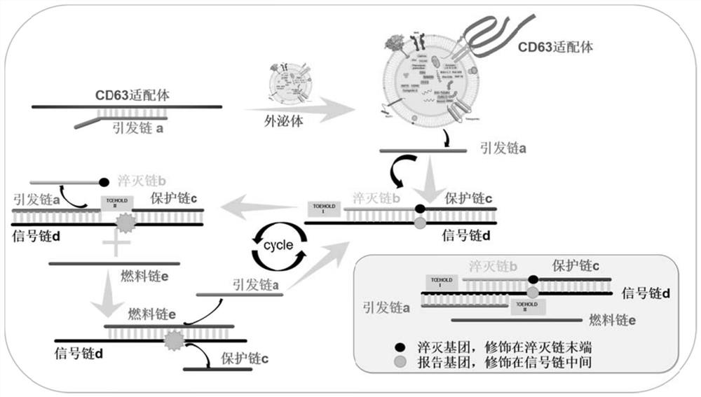
[0062] DNA molecular nanomachines for the quantification of exosomes in tumor cell culture supernatants, including:

[0063] The recognition probe is a double-stranded DNA structure formed by hybridizing the priming strand a with the nucleic acid aptamer (Apt-CD63) sequence of CD63. Preparation of the recognition probe: Add the nucleic acid aptamer and priming strand a to a certain volume of ligation reaction buffer at a final concentration ratio of 1:1, denature at 95°C for 5 minutes to open the local secondary structure of the DNA strand, and quickly Cool down to room temperature, so that the two oligonucleotide chains hybridize into a recognition probe, and store at 4°C for future use.

[0064] The signal probe is a double-stranded DNA structure formed by the hybridization of two short DNA strands (quencher strand b and protection strand c modified at the end of BHQ2) and a long DNA strand (reporter strand modified in the middle of Cy3). After the signal probe self-assembl...

Embodiment 2

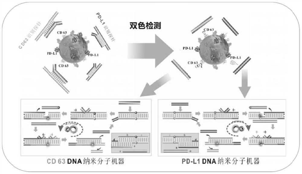
[0073] DNA nanomolecular machines for two-color phenotyping of exosomes (CD63 and PD-L1) in tumor cell culture supernatants, including:

[0074] Recognition probe: CD63 recognition probe is initiated by strand a CD63 Aptamers to CD63 (Apt CD63 ) double-stranded DNA structure formed by partial hybridization of sequences. The PD-L1 recognition probe is initiated by a PD-L1 In the nucleic acid aptamer (Apt PD-L1 ) double-stranded DNA structure formed by partial hybridization of sequences. The preparation method of the recognition probe is the same as that in Example 1.

[0075] Signal probe: The CD63 signal probe is a quenched chain b modified by two short DNA strands (BHQ2 ends) CD63 and protection chain c CD63 ) and a long DNA strand (reporter strand modified in the middle of Cy3) to form a double-stranded DNA structure. After the signal probe self-assembled successfully, the Cy3 and BHQ2 groups modified internally caused the fluorescence of Cy3 to be quenched by BHQ2 du...


PUM


Abstract

The invention relates to a DNA nano-molecule machine for exosome and surface protein analysis and application. The DNA nano-molecule machine comprises an identification probe generating response to asurface marker of an exosome, a signal probe containing two Toehold regions and a fuel probe capable of driving cycling of a strand displacement reaction. The exosome is subjected to polyvalent conjugation with a specific aptamer in the identification probe through the surface marker of the exosome, so as to release an initiating strand in the identification probe; the initiating strand invades the signal probe and drives branches of the strand to migrate and release a quenching strand through being conjugated with a Toehold region I, a fluorescence reporter group is liberated, and a concealedToehold region II is exposed; and the fuel probe starts up a Toehold-mediated strand displacement cyclic reaction through being conjugated with the Toehold region II, and continuous branch migrationand polyvalent recovery occur. Supersensitive quantitative detection on the exosome can be achieved; bi-color or multicolor analysis on two or more kinds of protein markers on the surface of the exosome can be achieved; and the operation is simple, the consumed time is short, the sample consumption is low, the sensitivity is high, and the specificity is good.

Description

technical field [0001] The invention belongs to the technical field of rapid detection, and in particular relates to a DNA nanomolecular machine driven by exosomes or surface proteins of exosomes, which can be used for qualitative and quantitative analysis of exosomes and surface proteins. It has a signal-response mechanism for multivalent cyclic amplification and can be used for ultrasensitive detection of tumor exosomes and exosomal phenotype molecules. Background technique [0002] In recent years, the liquid biopsy technology of precision medicine has been widely used in the early screening of tumors, auxiliary diagnosis, efficacy monitoring and drug efficacy prediction. Compared with traditional tissue biopsy, liquid biopsy has unique advantages such as non-invasiveness, real-time dynamic detection, overcoming tumor heterogeneity, and providing comprehensive detection information. [0003] In current clinical research, liquid biopsy techniques mainly include detection ...

Claims


Application Information

Patent Timeline
IPC IPC(8): C12Q1/6825C12Q1/682G01N33/569G01N33/68
CPCC12Q1/6825C12Q1/682G01N33/56966G01N33/68G01N2333/70596G01N2333/70532C12Q2525/205C12Q2563/107C12Q2565/1015Y02A50/30
Inventor 杨帆张国军金丹
Owner HUBEI UNIV OF CHINESE MEDICINE
Why Eureka
Try Eureka