Preparation method of culture substrate for batch production of 3D cell spheres

A culture substrate and mass production technology, applied in the field of biomedical material preparation, can solve the problems of expensive 3D cell spheroid culture substrate, large differences in cell spheroid size, unfavorable cell spheroid growth, etc., to improve accuracy and repeatability , the experimental cost reduction, the effect of uniform diameter and structure

Active Publication Date: 2021-03-02
XUZHOU MEDICAL UNIV
View PDF4 Cites 1 Cited by
  • Summary
  • Abstract
  • Description
  • Claims
  • Application Information

AI Technical Summary

Problems solved by technology

However, these methods have certain limitations, such as the contact between the two sides and the tube wall is not conducive to the growth of cell spheres, the size of the resulting cell spheres varies greatly, the large-scale preparation is limited, the collection of cell spheres is difficult, or more advanced equipment is required.
Moreover, the 3D cell spheroid culture substrate produced on the market is expensive, so it is of great significance to develop a practical and inexpensive batch culture 3D cell spheroid substrate

Method used


Image

  • Preparation method of culture substrate for batch production of 3D cell spheres
  • Preparation method of culture substrate for batch production of 3D cell spheres

Examples


Embodiment 1

[0027] Effect of Gelatin Concentration

[0028] 1. Obtaining of gelatin microspheres: gelatin particles were dissolved in ultrapure water at 60°C in a water bath, and gelatin solutions with concentrations of 5%, 8%, and 10% (wt / v, g / mL) were prepared respectively. Collect gelatin microspheres with a microfluidic device, by controlling the flow rate of the aqueous phase solution (gelatin solution) to be 5ml / h, the flow rate of the organic phase (based on toluene, adding 3wt% Span80 as a surfactant) flow rate is 40ml / h, and the capillary inner diameter 0.85mm is used as the organic phase channel, and a syringe needle (0.45mm inner diameter) is used as the aqueous phase solution channel inside the capillary to obtain gelatin microspheres, which are collected in methanol solution for subsequent use;

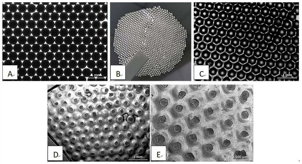
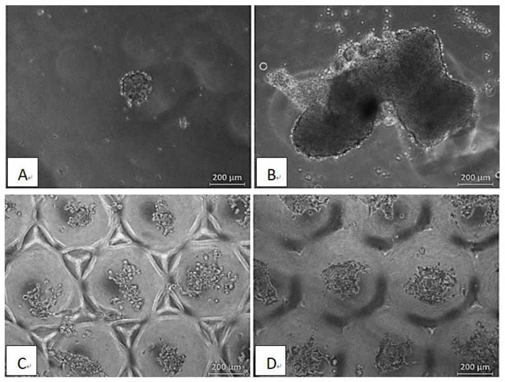
[0029] 2. Preparation of gelatin template: drop gelatin microspheres into a watch glass from the edge, slightly vibrate the watch glass to arrange the gelatin microspheres in a singl...

Embodiment 2

[0036] Influence of Channel Diameter of Aqueous Solution

[0037] This example is basically the same as Example 1, except that the concentration of gelatin is 8%, and the diameters of the channels of the aqueous phase solution are controlled to be 0.3mm and 0.45mm respectively.

[0038] The average diameters of the gelatin microspheres prepared with the channel diameters of 0.3 mm and 0.45 mm in the aqueous phase were 300 μm and 500 μm, respectively. Gelatin microspheres with uniform diameters can be obtained at both flow rates, and the diameters are slightly different. Based on the requirements for subsequent 3D cell sphere culture, the diameter of the aqueous phase channel of 0.45 mm is selected.

Embodiment 3

[0040] Influence of Channel Diameter of Organic Phase

[0041] This example is basically the same as Example 1, except that the gelatin concentration is 8%, and the channel diameters of the organic phase are controlled to be 0.85mm and 1.2mm respectively.

[0042] The average diameters of the gelatin microspheres prepared with organic phase channel diameters of 0.85 mm and 1.2 mm were 500 μm and 900 μm, respectively. It can be seen that increasing the channel diameter of the organic phase can obtain gelatin microspheres with larger sizes, indicating that the present invention can adjust the size of the obtained gelatin microspheres by changing the inner diameters of the organic phase and aqueous phase channels, and further affect the size of the subsequent 3D cell spheres.


PUM

PropertyMeasurementUnit
diameteraaaaaaaaaa
diameteraaaaaaaaaa
diameteraaaaaaaaaa

Abstract

The invention discloses a preparation method of a culture substrate for batch production of 3D cell spheres. The method comprises the steps: preparing gelatin microspheres with uniform diameters by adopting a microfluidic technology, obtaining a single-layer arrangement template of the gelatin microspheres through a watch glass, heating to tightly arrange the gelatin microspheres, pouring PDMS, curing, removing the gelatin template by adopting a water bath method to obtain a PDMS porous microarray substrate, and finally soaking the substrate in an F127 solution to obtain the culture substrateof the 3D cell spheres. PDMS porous microarray substrates with different pore diameters can be obtained by changing the diameters of the gelatin microspheres, so that 3D cell spheres with different sizes can be obtained. The method is simple and easy to implement, controllable in size, low in cost and suitable for batch production of the 3D cell spheres.

Description

technical field [0001] The invention relates to a method for preparing a culture substrate for batch production of 3D cell spheroids, and belongs to the technical field of preparation of biomedical materials. Background technique [0002] Traditional 2D monolayer cell culture is difficult to properly reflect the in vivo growth environment of cells, which may cause distortion of cell structure and tissue function. In contrast, the morphology and function of cells cultured in 3D are closer to the real growth conditions in animals. In addition, in the process of tumor drug screening, traditional 2D tumor cell culture is difficult to reflect the real situation in vivo, resulting in the failure of effective drugs tested in vitro when tested in vivo. Therefore, it is crucial to culture 3D tumor cell spheroids in vitro to mimic tumor conditions in vivo. [0003] The existing preparation methods of 3D cell spheroids include photolithography multi-well plate, forced floating, agita...

Claims


Application Information

Patent Timeline
Patent Type & Authority Applications(China)
IPC IPC(8): C12N5/00C08J9/42C08L83/04
CPCC12N5/0068C08J9/42C12N2513/00C12N2533/30C08J2383/04C08J2471/02
Inventor 吕兰欣梁琪沈红先李猛赵文轩金春燕王晶胡书群
Owner XUZHOU MEDICAL UNIV
Why Eureka
Try Eureka