Separation and enrichment method for quickly extracting tissue extracellular vesicles
A technology for separation, enrichment, and tissue cells, which is applied in the biological field, can solve the problems of cumbersome operation, long extraction time of tissue extracellular vesicles, and low purity, and achieve the effects of simplified steps, low pollution of soluble impurities, and high purity
- Summary
- Abstract
- Description
- Claims
- Application Information
AI Technical Summary
Problems solved by technology
Method used
Image
Examples
Embodiment 1
[0040] Embodiment 1 Enzymolysis process optimization experiment
[0041] In the early stage of the experiment, papain, collagenase + DNase I, and tissue-digesting enzyme were used to dissociate mouse heart tissue, and the optimal tissue-digesting enzyme was selected by comparing the experimental results, as follows:
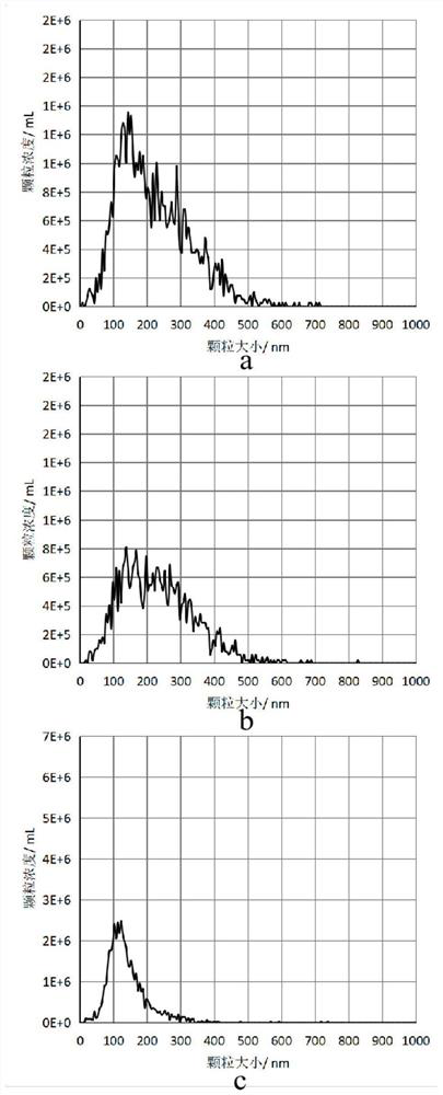
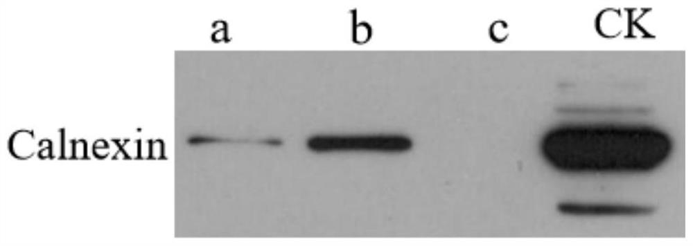
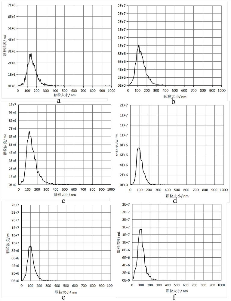
[0042] 1. Papain dissolving scheme: the dosage of papain (Worthington-biochemical, LK003178) is 15-30U, and the papain solution used is to dissolve papain with Earle's balanced salt solution (EBSS buffer), and then add Hibernate E medium to obtain wherein, the volume ratio of Earle's balanced salt solution to Hibernate E medium is 1:3, and the amount of papain solution is about 3mL; the extracellular vesicle size analysis diagram of papain is shown in figure 1 As shown in a, the WB of Calnexin protein is shown as figure 2 As shown in a, the mass of mouse heart tissue and the total amount of BCA protein are shown in Table 1.
[0043]2. Collagenase D and DNase I...
Embodiment 2
[0048] Example 2 Enrichment method for rapid extraction of extracellular vesicles from mouse heart tissue
[0049] The method for enriching extracellular vesicles in mouse heart tissue, the specific steps are as follows:
[0050] 1. Cut the cryogenically frozen (-80°C) tissue into 1-2 mm with a blade 3 size;
[0051] 2. The tissue slices were transferred to a centrifuge tube containing 2.5 mL of tissue-digesting enzyme dissociation solution, wherein the preparation of the tissue-digesting enzyme was the same as in Example 1;
[0052] 3. Put the above mixture into a 37°C water bath, mix it every 5 minutes, and dissociate for 30 minutes to obtain a tissue dissociation solution, and then filter the above tissue dissociation solution with a 70 μm filter membrane into a new centrifuge tube to obtain a tissue cell suspension;
[0053] 4. Add 25uL protease and phosphatase inhibitor mixture to the tissue cell suspension;
[0054] 5. Centrifuge the mixture in step 4 at 300-600 g fo...
Embodiment 3
[0063] Example 3 Enrichment method for rapid extraction of extracellular vesicles from mouse liver tissue
[0064] The method for enriching extracellular vesicles in mouse liver tissue, the specific steps are as follows:
[0065] 1. Cut the cryogenically frozen (-80°C) tissue into 1-2 mm with a blade 3 size;
[0066] 2. The tissue slices were transferred to a centrifuge tube containing 3mL of tissue-digesting enzyme dissociation solution, wherein the preparation of tissue-digesting enzyme was the same as in Example 1;
[0067] 3. Put the above mixture into a 37°C water bath, mix it every 5 minutes, and dissociate for 20 minutes to obtain a tissue dissociation solution, and then filter the above tissue dissociation solution into a new centrifuge tube with a 70 μm filter membrane to obtain a tissue cell suspension;
[0068] 4. Add 30uL protease and phosphatase inhibitor mixture to the tissue cell suspension;
[0069] 5. Centrifuge the mixture in step 4 at 300-600g for 10min ...
PUM
| Property | Measurement | Unit |
|---|---|---|
| Particle size | aaaaa | aaaaa |
| Particle size | aaaaa | aaaaa |
Abstract
Description
Claims
Application Information
 - R&D
- Intellectual Property
- Life Sciences
- Materials
- Tech Scout
- Unparalleled Data Quality
- Higher Quality Content
- 60% Fewer Hallucinations
Browse by: Latest US Patents, China's latest patents, Technical Efficacy Thesaurus, Application Domain, Technology Topic, Popular Technical Reports.
© 2025 PatSnap. All rights reserved.Legal|Privacy policy|Modern Slavery Act Transparency Statement|Sitemap|About US| Contact US: help@patsnap.com


