Three-dimensional panoramic tomography ultrasonic device

A tomographic and panoramic technology, applied in the field of biomedical imaging, can solve the problems of reducing the repeatability of experiments, data consistency, unable to guarantee image consistency, limiting the field of view of single imaging, etc., to overcome the small field of view of high-resolution ultrasound, improve Reproducible, impact-reducing effects

Inactive Publication Date: 2021-04-09
UNIV OF SCI & TECH OF CHINA
View PDF7 Cites 0 Cited by
  • Summary
  • Abstract
  • Description
  • Claims
  • Application Information

AI Technical Summary

Problems solved by technology

However, the attenuation of ultrasound in tissue is closely related to frequency, and the increase of ultrasound frequency will greatly reduce the penetration depth in tissue, thus limiting the field of view of a single imaging
At the same time, ultrasound imaging usually requires the operator to manually control the ultrasound probe to contact the skin of the imaged person for imaging, so that the tissue of the imaged person is squeezed, and this squeeze cannot be guaranteed to be the same for each imaging, so even the same operation Even if the same object is imaged multiple times, the consistency of the images obtained by different samplings cannot be guaranteed, which reduces the repeatability of the experiment and the consistency of the data.

Method used


Image

  • Three-dimensional panoramic tomography ultrasonic device
  • Three-dimensional panoramic tomography ultrasonic device
  • Three-dimensional panoramic tomography ultrasonic device

Examples


Embodiment 1

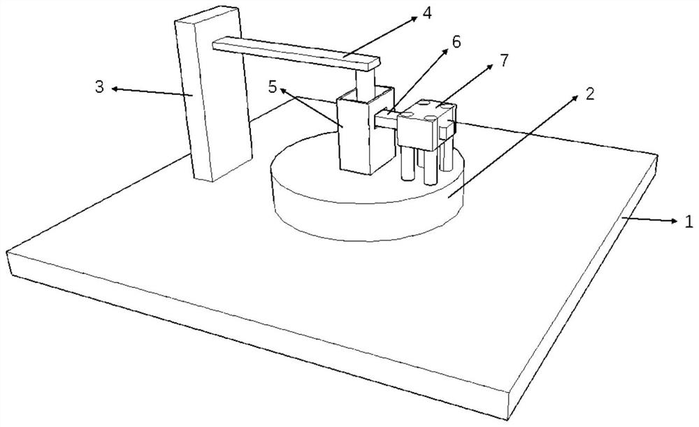
[0027] Embodiment 1: see again figure 1 , at this time the imaging plane of the ultrasonic probe 6 is parallel to the table surface of the rotary translation stage 2 and perpendicular to the rotation axis of the rotary translation platform 2 . After selecting a small animal for the experiment, first anesthetize the small animal, and then remove the hair of the imaging site to avoid the attenuation of the ultrasonic signal by the hair. After the pretreatment of the small animal is completed, place and fix the small animal on the L-shaped fixture 4, then put the cylinder of the L-shaped fixture 4 into the water tank 5, and adjust the lifting platform 3 so that the ultrasound connected to the fixture 7 The imaging plane of the probe 6 is aligned with the uppermost layer of the small animal imaging area. At this time, smear the ultrasonic coupling agent on the ultrasonic probe 6, put a dielectric film (such as a plastic bag, etc.) in the water tank 5, and add water to the water t...

Embodiment 2

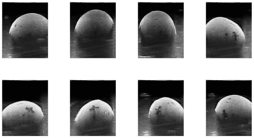
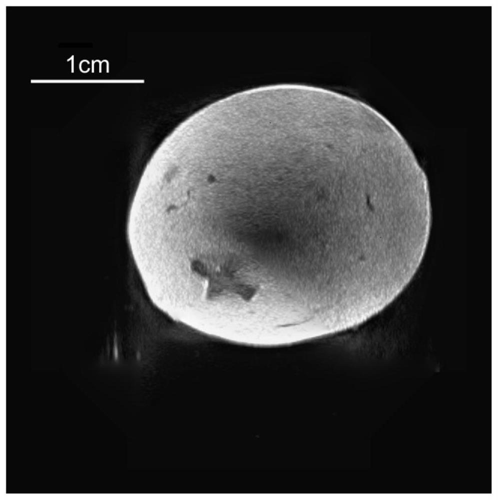
[0028] Embodiment 2: see illustration figure 1, at this time the imaging plane of the ultrasonic probe 6 is perpendicular to the table top of the rotary translation stage 2 , and the rotation axis of the rotary translation platform 2 is within the imaging plane of the ultrasonic probe 6 . The small animals were anesthetized, and the placement was the same as in Example 1. After the preparation is completed, the first frame of image is collected, and then the rotating stage 2 is operated to rotate 1 degree clockwise to collect the next frame of image, and this operation is repeated until the 360-degree image is collected. Since each image acquisition is a vertical section of a relatively high height of the imaging area, the data acquisition of the imaging area can usually be completed without raising and lowering the small animal. After the collection is completed, the data is input into the computer for subsequent processing. Rotate each collected image by 0, 1, 2, ... 359 i...


PUM


Abstract

The invention discloses a three-dimensional panoramic tomography ultrasonic device. The three-dimensional panoramic tomography ultrasonic device comprises a standard bread board, a motion control module, a fixing module and an imaging module, wherein the motion control module is positioned on the standard bread board and is fixedly connected with the standard bread board; the imaging module and the fixing module are fixed on the motion control module; during imaging, the motion control module drives the imaging module to rotate around a rotating shaft of the motion control module; and during imaging, the motion control module drives the fixing module to ascend and descend. An ultrasonic probe is fixed on a rotary moving table, and imaging repeatability is improved. A fixed imaging region is provided through a water tank, non-contact imaging is realized, and influences of an operator on a result are reduced. Complete data acquisition of a single fault is acquired through the rotary moving table, a panoramic image is obtained through an image fusion algorithm, and the problem that the visual field of existing high-resolution ultrasound is small is solved. Different fault images are acquired through an electric lifting table, and three-dimensional panoramic images of small animals are acquired.

Description

technical field [0001] The invention belongs to the technical field of biomedical imaging, and in particular relates to a three-dimensional panoramic tomographic ultrasonic device. Background technique [0002] Ultrasound is a kind of mechanical wave that propagates in the form of longitudinal waves in biological tissues. Ultrasonic imaging can be realized by transmitting and receiving returned ultrasonic waves through a transducer. Ultrasound technology for small animals generally uses high-frequency ultrasonic waves above 15MHz to achieve higher resolution and is used to detect superficial tissue structures or lesion information in small animals. It plays an important role in the preclinical research of pet medicine and disease. However, the attenuation of ultrasound in tissue is closely related to frequency, and the increase of ultrasound frequency will greatly reduce the penetration depth in tissue, thus limiting the field of view of a single imaging. At the same time,...

Claims


Application Information

Patent Timeline
Patent Type & Authority Applications(China)
IPC IPC(8): A61B8/08A61B8/13A61B8/00
CPCA61B8/085A61B8/13A61B8/4444A61B8/4281A61B8/483A61B2503/40
Inventor 杨益东陈家豪
Owner UNIV OF SCI & TECH OF CHINA
Why Eureka
Try Eureka