Method for detecting in-vitro inhibition of lymphocyte proliferation of human amniotic epithelial cells
A lymphocyte proliferation and epithelial cell technology, applied in the field of lymphocyte proliferation inhibition, can solve the problems of slow reproduction, large data fluctuation, and difference in the quality of human amniotic cells.
- Summary
- Abstract
- Description
- Claims
- Application Information
AI Technical Summary
Problems solved by technology
Method used
Image
Examples
Embodiment 1
[0061]Example 1 of the present invention provides a method of detecting a proliferative inhibition of lymphocytes in vitro in human amniotic membrane epithelial cells, which is as specifically as follows:
[0062](1) Cascardine epithelial cell vaccination: Take 400 g of cell suspension, centrifuge for 5 min, to the supernatant, add 10% by weight of the gibu blood-free GIBCO 1640 medium heavy, sampling count; adjust the cell concentration to 2 × 10⁵ / ml; the adjusted cell suspension was inoculated into 3 24-well culture plates, and 4 holes were inoculated each, and 12 holes were inoculated with 500 μl per well. Place the inoculated cell plate in a carbon dioxide incubator (CO2The concentration is set to: 5.0 vol%, the temperature is set to: 37.0 ° C), cultured for 20 h;
[0063](2) Subside each well medium in step (1), 500 μL of GIBCO 1640 medium containing 8 μg / ml of silk serum Ceramine Ceramine Ceramine, in the carbon dioxide incubator (CO)2The concentration is set to: 5.0 vol%, the te...
Embodiment 2
[0067]Example 2 of the present invention provides a method of detecting a proliferative inhibition of lymphocytes in vitro in human amniocenteular epithelial cells, which is specifically:
[0068](1) Cascardine epithelial cell vaccination: Take 400 g of cell suspension, centrifuge for 5 min, to the supernatant, add 10% by weight of the gibu blood-free GIBCO 1640 medium heavy, sampling count; adjust the cell concentration to 2 × 10⁵ / ml; the adjusted cell suspension was inoculated into 3 24-well culture plates, and 4 holes were inoculated each, and 12 holes were inoculated with 500 μl per well. Place the inoculated cell plate in a carbon dioxide incubator (CO2The concentration is set to: 5.0 vol%, the temperature is set to: 37.0 ° C), cultured for 20 h;
[0069](2) Subside each well medium in step (1), 500 μl of GIBCO 1640 medium containing 10 wt% fetal bovine serum containing 12 μg / ml of keteng serum is placed in a carbon dioxide incubator (CO) each of the GIBCO 1640 medium containing 1...
Embodiment 3
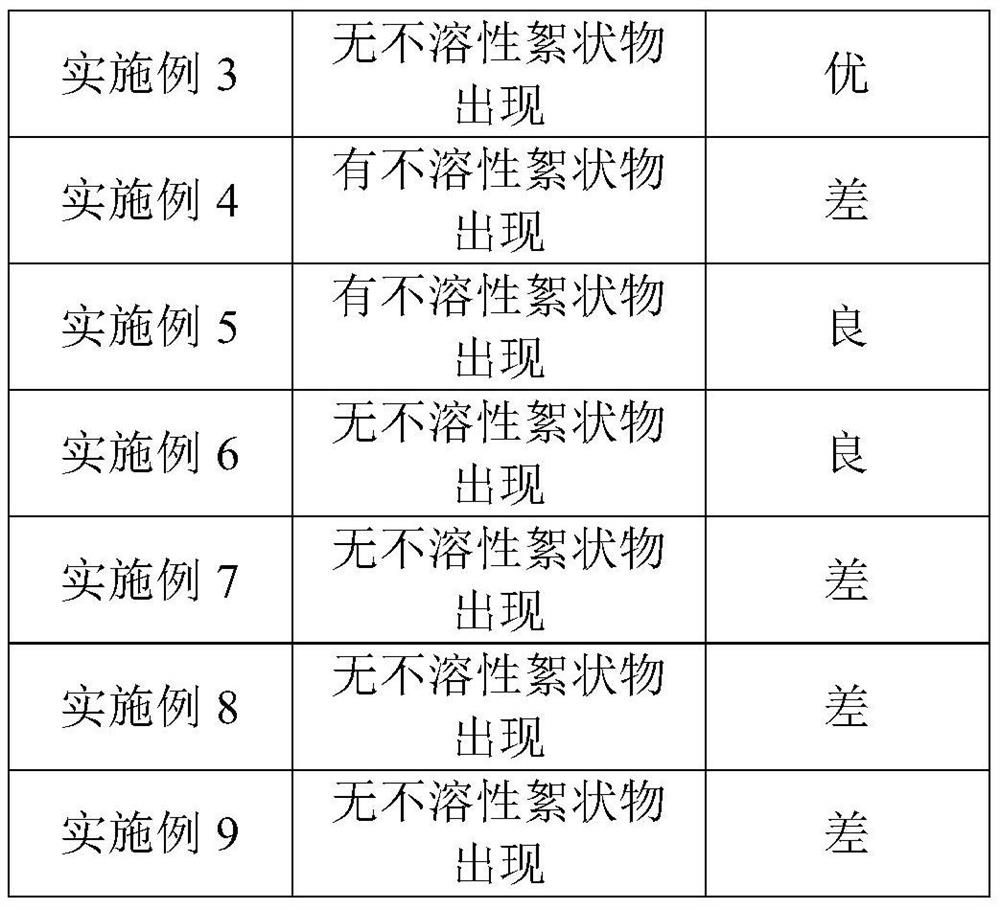
[0073]Example 3 of the present invention provides a method of detecting a proliferative method of lamb epithelial cells inhibiting lymphocytes, which is specifically:
[0074](1) Cascardine epithelial cell vaccination: Take 400 g of cell suspension, centrifuge for 5 min, to the supernatant, add 10% by weight of the gibu blood-free GIBCO 1640 medium heavy, sampling count; adjust the cell concentration to 2 × 10⁵ / ml; the adjusted cell suspension was inoculated into 3 24-well culture plates, and 4 holes were inoculated each, and 12 holes were inoculated with 500 μl per well. Place the inoculated cell plate in a carbon dioxide incubator (CO2The concentration is set to: 5.0 vol%, the temperature is set to: 37.0 ° C), cultured for 20 h;
[0075](2) Subside each well medium in step (1), 500 μl of GIBCO 1640 medium containing 10 wt% fetal bovine serum containing 10 wt% fetal bovine serum is placed in a carbon dioxide incubator for each GIBCO 1640 medium containing 10 wt% fetal bovine serum Co.2T...
PUM
 Abstract
Description
Claims
Application Information
 - R&D
- Intellectual Property
- Life Sciences
- Materials
- Tech Scout
- Unparalleled Data Quality
- Higher Quality Content
- 60% Fewer Hallucinations
Browse by: Latest US Patents, China's latest patents, Technical Efficacy Thesaurus, Application Domain, Technology Topic, Popular Technical Reports.
© 2025 PatSnap. All rights reserved.Legal|Privacy policy|Modern Slavery Act Transparency Statement|Sitemap|About US| Contact US: help@patsnap.com


