Extracellular vesicle enrichment detection method

A detection method and enrichment technology, applied in the field of medical detection, can solve the problems of low purity and recovery, poor effect, low efficiency, etc.

Pending Publication Date: 2021-09-07
CHONGQING UNIV
View PDF7 Cites 2 Cited by
  • Summary
  • Abstract
  • Description
  • Claims
  • Application Information

AI Technical Summary

Problems solved by technology

However, during the ultrafiltration process of ultrafiltration, exosomes may block the filter pores, resulting in shortened membrane life and low separation efficiency. In addition, exosome membranes will also adhere to each other, resulting in low separation yield and even False detection results occur; polyethylene glycol in the PEG-base precipitation method can compete with free water molecules, so that less soluble molecules or exosomes are precipitated from the solution. There are also some problems in this method: such as exosomes The purity and recovery rate of exosomes are low, and mechanical force or chemical additives can damage exosomes, etc.; while the magnetic bead immunoassay method can obtain high-purity exosomes due to the high specificity of immunoaffinity technology without affecting The advantages of complete exosome morphology are the preferred method for enriching and characterizing unique exosomes, but this method is inefficient, and the biological activity of exosome contents is easily affected by pH and salt concentration, which is not conducive to the downstream experiments
[0005] DNA origami technology is to repeatedly fold long-chain DNA to construct various tiny 3D structures, including micro-biosensors and drug delivery containers. At present, this technology has been applied to exosome detection, but the structure and construction rules are complicated. It is difficult to design and visualize the double-helix chain of DNA molecules bent into a specific shape. The existing technology focuses more on the labeling and detection of exosomes, and its origami structure is not effective in the enrichment and separation of exosomes.

Method used


Image

  • Extracellular vesicle enrichment detection method
  • Extracellular vesicle enrichment detection method
  • Extracellular vesicle enrichment detection method

Examples


Embodiment 1

[0044] Example 1 A kind of extracellular vesicle enrichment detection method

[0045] Experimental materials: DNA nucleic acid manufacturer is Shanghai Sangong, magnetic bead manufacturer is Thermo Fisher Scientific, antibody manufacturer is Proteintech, six-well plate manufacturer is Jingan Biological J06001;

[0046] TM buffer: containing 20mM Tris and 50mM MgCl.

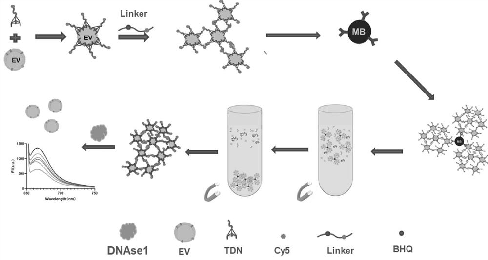
[0047] figure 1 For the experimental schematic diagram of the present embodiment, the method of the present embodiment comprises the following steps carried out in sequence:

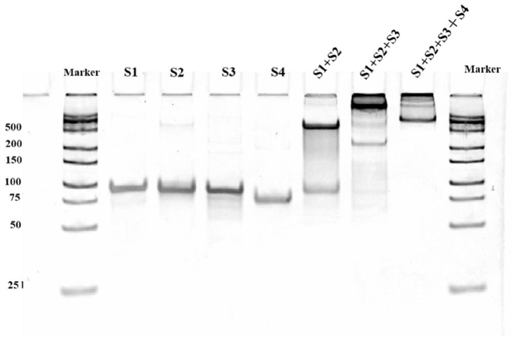
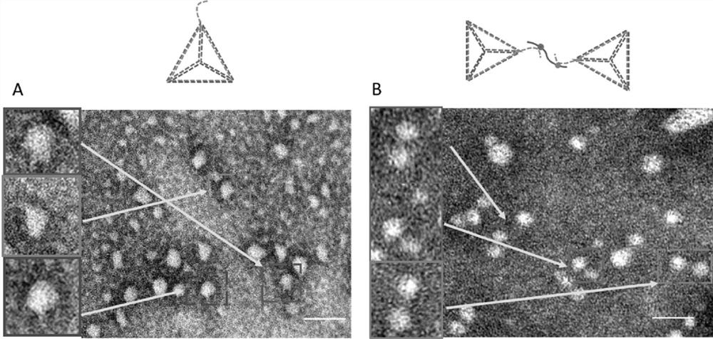
[0048] S1: DNA tetrahedron synthesis:

[0049] Design four DNA oligonucleotide chains L1, L2, L3 and L4, among which L1, L2 and L3 are modified with CD63 aptamers to assemble into the specific bottom of DNA tetrahedron, and the top of L4 has a binding site for the link chain Linker point, to be combined to form a protruding free chain, which acts as a link;

[0050] The DNA sequences involved are shown in Table 1;

[0051] The four DNA ...

Embodiment 2

[0071] Example 2 A kind of extracellular vesicle enrichment detection method

[0072] Example 2 is a method for detecting the enrichment of extracellular vesicles, which is basically the same as Example 1, except that step S3 is different, and other steps are the same. Step S3 of this embodiment is specifically:

[0073] S3: Extract exosomes:

[0074] Take the suspended cells, collect the cells by centrifugation, bounce the cells vigorously, add the lysate according to the ratio of 100-200 μL lysate per well of the six-well plate, and flick to fully lyse the cells until there is no obvious cell precipitation, remove Wash the culture medium once with PBS and normal saline to remove impurities, disperse the cells, add the lysate according to the ratio of adding 100mL (200uL for 6cm petri dish) to each well of the six-well plate, fully lyse on ice for 25min, and then centrifuge at 10000g for 3min. The extracted supernatant, which contains the exosomes to be enriched, can be subj...

Embodiment 3-5

[0075] Example 3-5 Extracellular vesicle enrichment detection method

[0076] Examples 3-5 are respectively a method for detecting extracellular vesicle enrichment, which are basically the same as in Example 1, except that the specific aptamers selected in Example 3-5 are CD9, CD91 and EPCAM in sequence specific aptamers.


PUM

PropertyMeasurementUnit
diameteraaaaaaaaaa

Abstract

The invention belongs to the technical field of medical detection, and discloses an extracellular vesicle enrichment detection method, which is characterized in that exosome enrichment detection is performed after DNA tetrahedron synthesis, magnetic bead combination and exosome extraction. According to the method, exosome surface specific protein is utilized, on the basis of a DNA origami aptamer and immunoaffinity magnetic bead space recognition dual recognition and capture method, a DNA fluorescent probe is further combined, so that rapid separation and capture of the exosome are realized, and the method can be used for quantitatively detecting the exosome. The invention is suitable for being used as a kit to realize enrichment, separation and detection of exosomes, and has very important significance in wide application in the fields of disease diagnosis, pathological research, new drug development and the like.

Description

technical field [0001] The invention belongs to the technical field of medical detection, and relates to an exosome enrichment detection method, in particular to an extracellular vesicle enrichment detection method. Background technique [0002] Exosomes refer to small vesicles secreted by cells, that is, extracellular vesicles, with a diameter of mostly between 30-150nm. Exosomes contain proteins, lipids, nucleic acids and other substances, which can be received by recipient cells to realize material transportation and information transmission between cells. Almost all biological cells can release exosomes in various states, and even some microorganisms can release exosomes for signal transmission. Exosomes contain many key proteins and special genetic materials, which play an important role in antigen presentation, immune regulation, tissue development, intercellular communication, and mediate tumor development and development in cell communication and epigenetic regulati...

Claims


Application Information

Patent Timeline
Patent Type & Authority Applications(China)
IPC IPC(8): G01N21/64G01N33/543
CPCG01N21/6486G01N33/54326
Inventor 罗阳于兴乐陈晓辉陈恒屹张亮亮胡孝林
Owner CHONGQING UNIV
Why Eureka
Try Eureka