Universal fluorescent biosensor for detecting ATP, glutathione and Fpg glycosylase
A biosensor and glycosylase technology, applied in the field of general-purpose fluorescent biosensors, can solve the problems of complicated instrument operation and expensive equipment, and achieve the effects of good repeatability, fast response and fast speed
- Summary
- Abstract
- Description
- Claims
- Application Information
AI Technical Summary
Problems solved by technology
Method used
Image
Examples
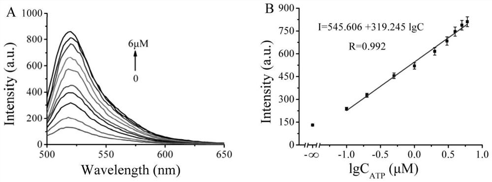
Embodiment 1
[0062] A preparation method of the optical biosensor of the present invention, comprising the following steps:
[0063] a. Put 5 µL sgRNA (50nM), 5 µL ATP block (50nM), 5 µL 1×TAE in a 1.5mL centrifuge tube, vortex and mix well, put it in a water bath at 95°C for 5 minutes, and then cool to room temperature ;
[0064] b. Take 5 µL Apt1 (100nM), 5 µL Apt2 (100nM), 5 µL Cas12a protein (100nM), 5 µL S chain (50nM), 5 µL 10×Buffer2.1, 1 µL FQ chain (1µM), 4 Add µL sterile water, 5 µL ATP (0 µM, 100nM, 200nM, 500nM, 1µM, 2µM, 3µM, 4µM, 5µM, 6µM) to the 15µL solution obtained in the previous step;
[0065] c. Place the centrifuge tube in a 37°C water bath for 90 minutes;
[0066] d. Place the centrifuge tube in a 65°C water bath for 10 minutes to terminate the reaction;
[0067] e. The excitation wavelength is 485nm, the emission wavelength is 520nm, the voltage is 670V, and the target to be tested is detected through the change of fluorescence.
[0068] see results figure 2 ,...
Embodiment 2
[0070] A preparation method of the optical biosensor of the present invention, comprising the following steps:
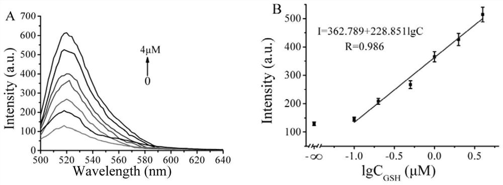
[0071] a. Put 5 µL sgRNA (50nM), 5 µL GSH block (50nM), 5 µL 1×TAE in a 1.5mL centrifuge tube, vortex and mix well, put it in a water bath at 95°C for 5 minutes, and then cool to room temperature ;
[0072] b. Take 5 µL Cas12a protein (100nM), 5 µL S chain (50nM), 5 µL 10×Buffer2.1, 1 µL FQ chain (1 µM), 14 µL sterile water, 5 µL GSH (0 µM, 100nM, 200nM , 500nM, 1µM, 2µM, 4µM), added to the 15µL solution obtained in the previous step;
[0073] c. Place the centrifuge tube in a 37°C water bath for 90 minutes;
[0074] d. Place the centrifuge tube in a 65°C water bath for 10 minutes to terminate the reaction;
[0075] e. The excitation wavelength is 485nm, the emission wavelength is 520nm, the voltage is 670V, and the target to be tested is detected through the change of fluorescence.
[0076] see results image 3 ,from image 3 It can be seen in A that the dete...
Embodiment 3
[0078] A preparation method of the optical biosensor of the present invention, comprising the following steps:
[0079] a. Put 5 µL sgRNA (50nM), 5 µL Fpg block (50nM), 5 µL 1×TAE in a 1.5mL centrifuge tube, vortex and mix well, put it in a water bath at 95°C for 5 minutes, and then cool to room temperature ;
[0080] b. Take 5 µL Cas12a protein (100nM), 5 µL S chain (50nM), 5 µL 10×Buffer2.1, 1 µL FQ chain (1 µM), 14 µL sterilized water, 5 µL Fpg (0 µM, 50nM, 100nM , 200nM, 500nM, 1µM), added to the 15µL solution obtained in the previous step;
[0081] c. Place the centrifuge tube in a 37°C water bath for 90 minutes;
[0082] d. Place the centrifuge tube in a 65°C water bath for 10 minutes to terminate the reaction;
[0083] e. The excitation wavelength is 485nm, the emission wavelength is 520nm, the voltage is 670V, and the target to be tested is detected through the change of fluorescence.
[0084] see results Figure 4 . from Figure 4 It can be seen in A that the d...
PUM
 Abstract
Description
Claims
Application Information
 - R&D
- Intellectual Property
- Life Sciences
- Materials
- Tech Scout
- Unparalleled Data Quality
- Higher Quality Content
- 60% Fewer Hallucinations
Browse by: Latest US Patents, China's latest patents, Technical Efficacy Thesaurus, Application Domain, Technology Topic, Popular Technical Reports.
© 2025 PatSnap. All rights reserved.Legal|Privacy policy|Modern Slavery Act Transparency Statement|Sitemap|About US| Contact US: help@patsnap.com


