Marker combination and kit for detecting tumor cells in body fluid sample
A detection kit and technology for tumor cells, applied in the field of marker combinations and kits for detection of tumor cells in body fluid samples, can solve the problems of false positives of tumor cells and achieve the effect of improving sensitivity and specificity
- Summary
- Abstract
- Description
- Claims
- Application Information
AI Technical Summary
Problems solved by technology
Method used
Image

Examples
Embodiment 1
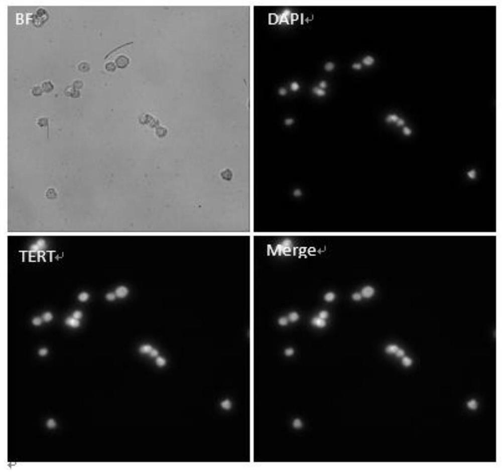
[0045] Example 1: Verification of Telomerase Expression in Tumor Cell Lines
[0046] (1) Centrifuge 1 ml of the MCF-7 cell line (500 g, 5 minutes), discard the supernatant, and add 2 mL of HBSS buffer to resuspend the cells;
[0047] (2) After the above resuspension is counted by a cell counter, take an appropriate amount and drop it on a glass slide, and let it stand for 10 minutes;
[0048] (3) Blot off the solution on the surface of the slide, add 100ul of TERT-FITC antibody with a concentration of 2ug / ml dropwise, and incubate at room temperature for 1h;
[0049] (4) After the incubation, wash the chip 3 times with PBS, drop 100ul of DAPI onto the glass slide, and incubate at room temperature for 10min;
[0050] (5) After the incubation, the chip was washed 3 times with PBS, and imaged with a high-speed fluorescence imaging device.
[0051] Imaging results such as figure 1 As shown, the images of cells with different fluorescence characteristics obtained by scanning sli...
Embodiment 2
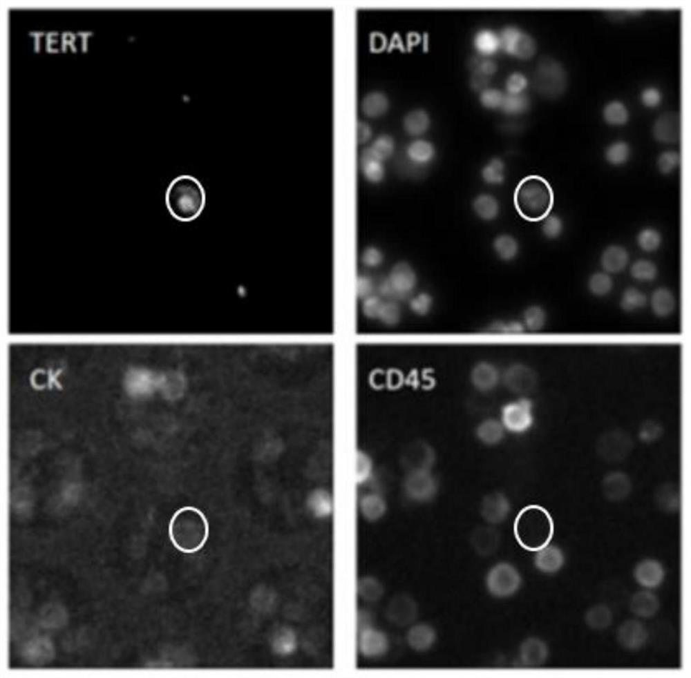
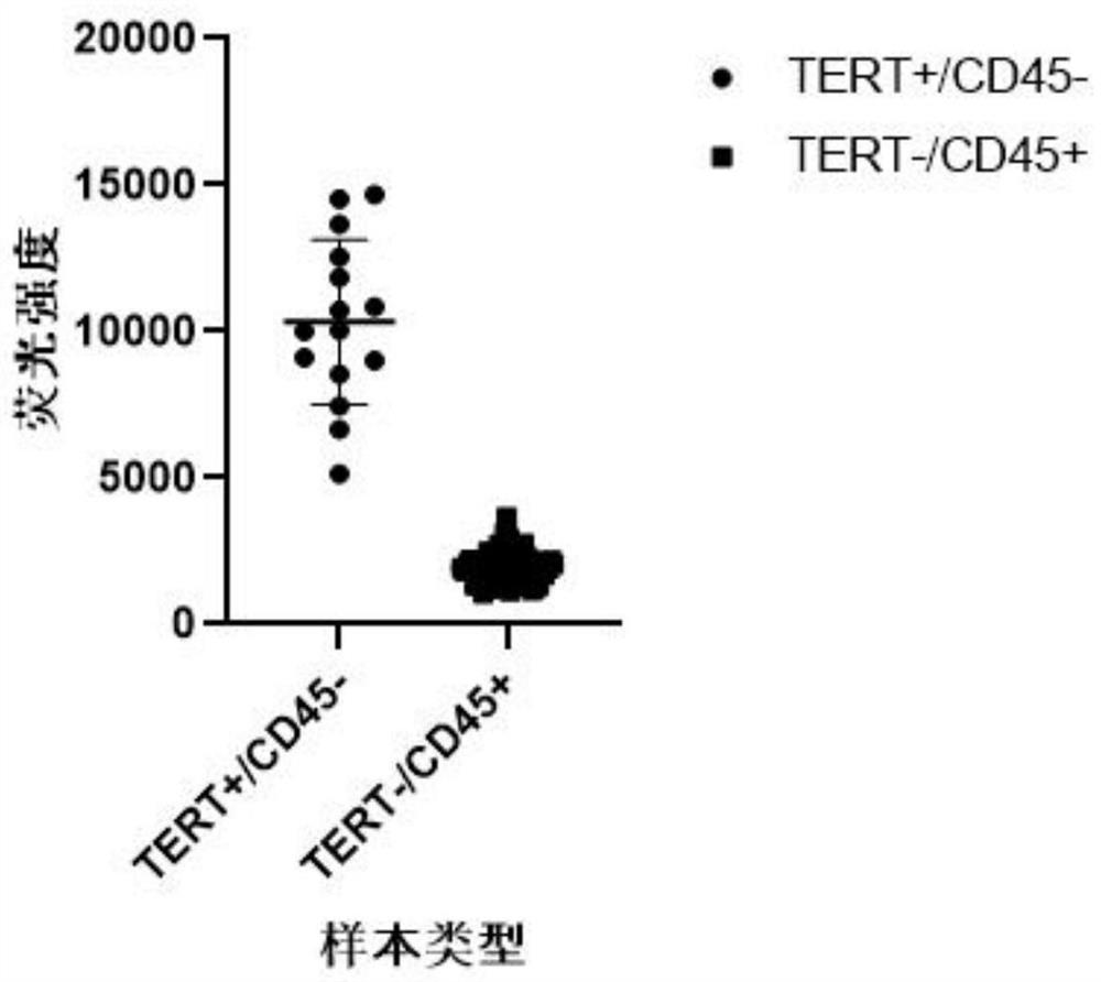
[0052] Example 2: Pleural effusion sample test of lung cancer patients
[0053] The testing method of the pleural effusion sample of lung cancer patient comprises the following steps:
[0054] (1) Centrifuge (500g, 5min) 20mL of pleural effusion from lung cancer patients to separate the cells, add 5mL red blood cell lysate (BD company) to lyse in the dark for 5min, centrifuge again (500g, 5min), discard the supernatant and use Hank's balanced salt Solution (HBSS) to resuspend and wash the cells, centrifuge (500g, 5min), discard the supernatant, add 2mL HBSS to resuspend the cells;
[0055] (2) After the above resuspension is counted by a cell counter, take an appropriate amount and drop it on a glass slide, and let it stand for 10 minutes;
[0056] (3) Absorb the solution on the surface of the slide, and drop 100ul antibody mixture onto the slide, wherein the antibody mixture includes: 2ug / ml FITC-labeled TERT antibody, PE-labeled 1ug / ml CK antibody and 2ug / ml ml of APC-labele...
Embodiment 3
[0061] Example 3: Urine Tests for Bladder Cancer Patients
[0062] The method of urine testing in patients with bladder cancer involves the following steps:
[0063] (1) Separate the cells from 20 mL of bladder cancer patient urine (500 g, 5 min), add 5 mL of erythrocyte lysate (BD company) to lyse in the dark for 5 min, centrifuge again (500 g, 5 min), discard the supernatant and equilibrate with Hank Resuspend and wash cells in saline solution (HBSS), centrifuge (500g, 5min), discard supernatant, add 2mL HBSS to resuspend cells;
[0064] (2) After the above resuspension is counted by a cell counter, take an appropriate amount and drop it on a glass slide, and let it stand for 10 minutes;
[0065] (3A) Absorb the solution on the surface of the slide, and drop 100ul antibody mixture onto the slide, wherein the antibody mixture includes: 2ug / ml FITC-labeled TERT antibody, 1ug / ml PE
[0066] Labeled CK antibody and 2ug / ml APC-labeled CD45 antibody, incubated at room temperatur...
PUM
 Abstract
Description
Claims
Application Information
 - R&D
- Intellectual Property
- Life Sciences
- Materials
- Tech Scout
- Unparalleled Data Quality
- Higher Quality Content
- 60% Fewer Hallucinations
Browse by: Latest US Patents, China's latest patents, Technical Efficacy Thesaurus, Application Domain, Technology Topic, Popular Technical Reports.
© 2025 PatSnap. All rights reserved.Legal|Privacy policy|Modern Slavery Act Transparency Statement|Sitemap|About US| Contact US: help@patsnap.com