Retina protective agent, retina protection method and application of protective agent
A protective agent, retinal technology, applied in the field of retina, can solve problems such as treatment termination and recurrence of lesions, and achieve the effects of low price, inhibiting oxidative damage, and delaying tissue aging.
- Summary
- Abstract
- Description
- Claims
- Application Information
AI Technical Summary
Problems solved by technology
Method used
Image
Examples
Embodiment 1
[0032] Example 1 (study on the protective effect of exogenous Ghrelin on the retinal tissue of db / db mice):
[0033] Proceed as follows:
[0034] Step 1: Grouping and treatment of type 2 diabetic mice db / db: Take 10-week-old db / db mice, and use the tail artery to measure blood glucose (blood glucose ≥ 16.6 mmol / L) to identify whether the diabetes model is successful. Animal groups: control group, model group and Ghrelin group. Ghrelin group: In the last two weeks of the model group, a subcutaneous micropump was given to continuously pump Ghrelin (5 μg / kg / d) for two weeks. .
[0035] Step 2: HE staining to examine the retinal structure: Dewax the retinal tissue with xylene, prepare paraffin sections, dehydrate with gradient alcohol, rinse repeatedly with distilled water, soak in hematoxylin staining solution for 10 minutes, stain with ammonia water and acid water, and soak in gradient alcohol solution 10 min, HE staining for 30 s, and taking pictures after mounting.
[0036...
Embodiment 2
[0040] Example 2 (HE staining to observe pathological and morphological changes of retinal tissue):
[0041] Three rats were randomly selected in each group, and after the left eyeball was removed, part of the retinal tissue was fixed with 4% paraformaldehyde for 2 hours, and the 5 μm retinal slices were placed on a glass slide, washed with gradient alcohol, stained with H&E, and light microscope (200 ×) Take the image.
[0042] Histopathological changes of S retina:
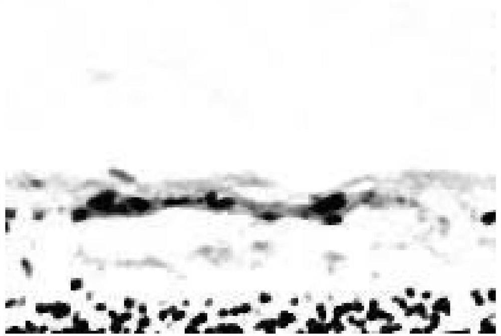
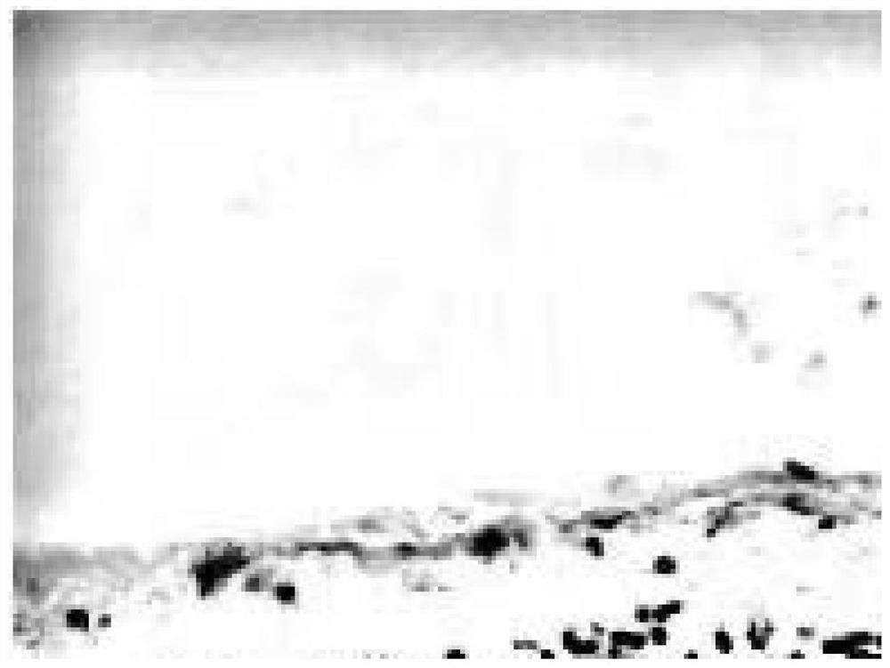
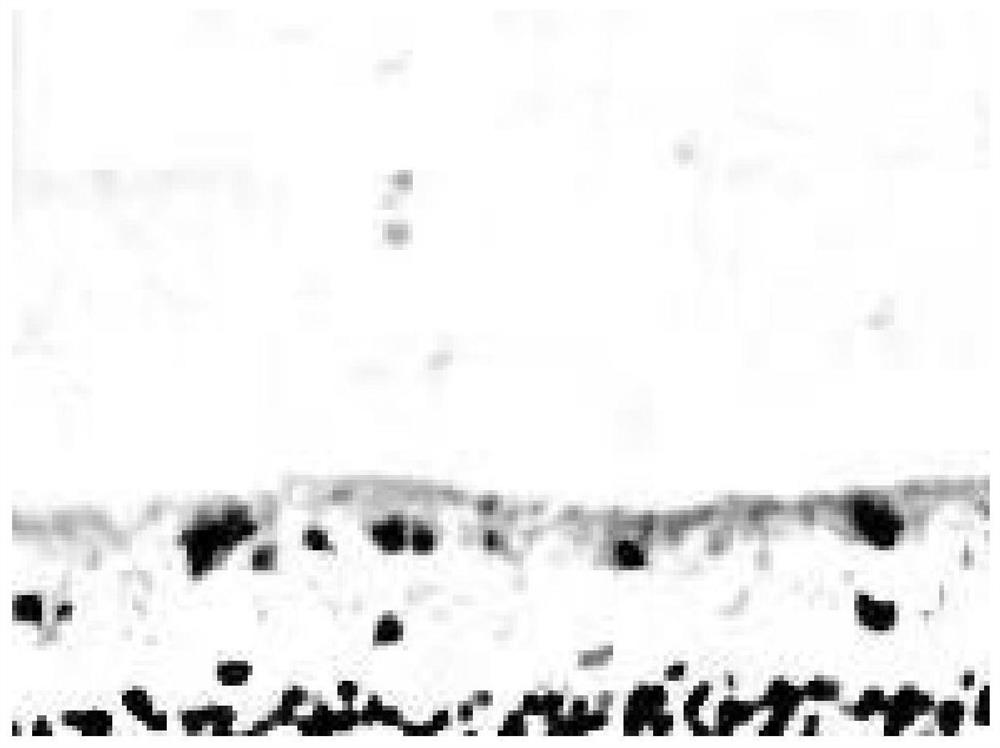
[0043] Such as Figure 1-3 as shown, figure 1 In the control group, the retinal tissue was complete and the structure was clear; figure 2 The thickness of the retina in the model group was significantly reduced, and the inner and outer nuclear layer cells were loosely arranged; image 3 The thickness of the retina in the ghrelin group was higher than that in the model group, and the structure was relatively regular.
Embodiment 3
[0044] Example 3 (TUNEL staining to detect rat retinal cell apoptosis):
[0045] After dewaxing, the retinal slices were dipped twice in xylene, dehydrated with alcohol gradients, added TUNEL reaction mixture, incubated in a wet box for 2 hours, dried and incorporated into DAB chromogenic solution, and observed under a microscope that the nuclei were brownish-yellow as TUNEL If it is positive, wash the section with phosphate buffer solution to stop color development, seal the section with neutral gum, and take the image with a light microscope (200×).
[0046] Results of retinal TUNEL staining:
[0047] Such as Figure 4-6 as shown, Figure 4 Dark brown TUNEL-positive cells were occasionally seen in the retinal photoreceptor cell layer of rats in the control group. Figure 5 The number of TUNEL-positive cells in the model group increased significantly. Figure 6 After ghrelin intervention, the number of positive cells stained by TUNEL decreased.
PUM
 Abstract
Description
Claims
Application Information
 - R&D Engineer
- R&D Manager
- IP Professional
- Industry Leading Data Capabilities
- Powerful AI technology
- Patent DNA Extraction
Browse by: Latest US Patents, China's latest patents, Technical Efficacy Thesaurus, Application Domain, Technology Topic, Popular Technical Reports.
© 2024 PatSnap. All rights reserved.Legal|Privacy policy|Modern Slavery Act Transparency Statement|Sitemap|About US| Contact US: help@patsnap.com


