Application of combination of tissue transparentizing method and histological method to detection of bacteria in tumor

A histological and transparent technology, applied in the field of detection, can solve the problem of misreading the number and location of bacteria, and achieve the effect of increasing accuracy and reducing false positives.

Pending Publication Date: 2021-11-12
ZHUJIANG HOSPITAL SOUTHERN MEDICAL UNIV
View PDF12 Cites 1 Cited by
  • Summary
  • Abstract
  • Description
  • Claims
  • Application Information

AI Technical Summary

Problems solved by technology

[0007] Accu-OPTIClear technology is to make the tissue optically transparent, thereby increasing the imaging depth and image contrast. However, the labeling of bacteria still depends on immunofluorescence (Immunofluorescence, IF), immunofluorescence (Immunofluorescence, IF) and in situ fluorescence hybridization (Fluorescence in situ hybridization). situ hybridization,

Method used


Image

  • Application of combination of tissue transparentizing method and histological method to detection of bacteria in tumor
  • Application of combination of tissue transparentizing method and histological method to detection of bacteria in tumor
  • Application of combination of tissue transparentizing method and histological method to detection of bacteria in tumor

Examples


Embodiment 1

[0055] A method for detecting bacteria in gliomas in combination with a tissue clearing method and an immunofluorescent labeling method, comprising the following steps:

[0056] 1) Prepare OPTIClear tissue clearing reagent (mix 20% (w / v) N-methylglucamine (Sigma-Aldrich #66930), 32% (w / v) isohexyl alcohol (Sigma-Aldrich #D2158) and 25% - Thiodiethanol (TDE) (Sigma-Aldrich #88559) solution was mixed, and the pH of the solution was adjusted to 7) with concentrated hydrochloric acid, and then sodium dodecyl sulfate (SDS) detergent was dissolved in OPTIClear tissue clearing reagent to obtain the concentration 4% SDS-OPTIClear mixture (SDS:OPTIClear=4:100);

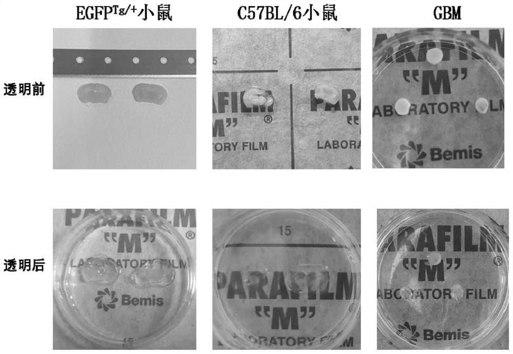
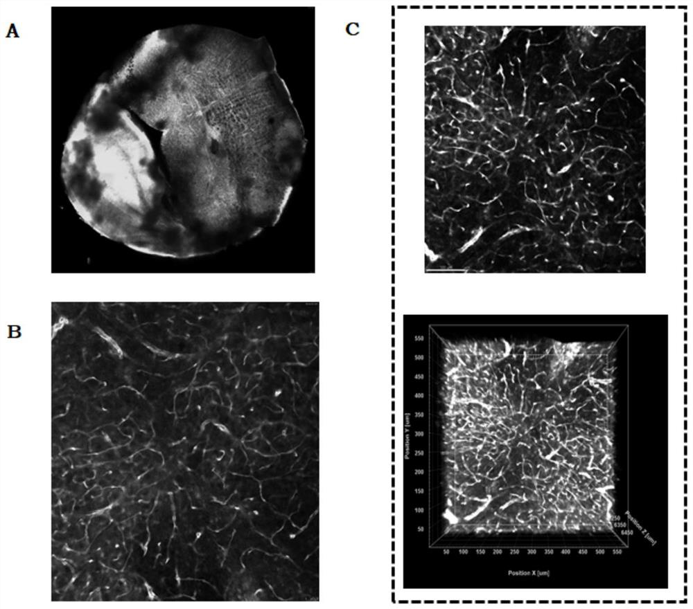
[0057] 2) Transfer the glioma tissue slices obtained above into a sterile EP tube, add SDS-OPTIClear mixed solution, so that the glioma tissue slices are soaked in the SDS-OPTIClear mixed solution, and the tissue slices are mixed with the SDS-OPTIClear mixed solution The volume ratio is 1:3, incubate at 37°C for 1-3 days, the...

Embodiment 2

[0071] An immunohistochemical method (IHC) is applied to the detection method of bacteria in human glioma, which comprises the following steps:

[0072] (1) Preparation of paraffin sections: The fixed human glioma samples were dehydrated and embedded in paraffin (dehydration: cherry blossom, VIPJ-JR; embedding: cherry blossom, TEC-5), and serial sections with a thickness of 4 μm were obtained (Leica , RM2245);

[0073] (2) Paraffin sections were dewaxed to water: put the sections in xylene I for 15 minutes, xylene II for 15 minutes, xylene III for 15 minutes, absolute ethanol I for 5 minutes, absolute ethanol II for 5 minutes, 85% alcohol for 5 minutes, and 75% alcohol for 5 minutes , and then washed with distilled water;

[0074] (3) Antigen retrieval: Place the tissue slices in a repair box filled with citric acid antigen retrieval buffer (PH6.0) for antigen retrieval in a microwave oven, heat for 8 minutes to boil, stop fire for 8 minutes, keep warm and then turn to medium...

Embodiment 3

[0083] An immunofluorescence method (IF) is applied to the detection method of bacteria in human glioma, which comprises the following steps:

[0084] (1) Preparation of tissue sections: The fixed human glioma samples were dehydrated and embedded in paraffin (dehydration: Cherry Blossom, VIPJ-JR; embedding: Sakura Blossom, TEC-5) to obtain serial sections with a thickness of 4 μm (Leica, RM2245), that is, paraffin section;

[0085] (2) Dewaxing and hydration of paraffin sections: put the sections in xylene I for 10 minutes, xylene II for 10 minutes, xylene III for 10 minutes, absolute ethanol for 5 minutes, 95% alcohol for 5 minutes, 85% alcohol for 5 minutes and 75% alcohol for 5 minutes, then Wash with distilled water 3 times, 5min each time;

[0086] (3) Antigen retrieval: Place the tissue slices in a repair box filled with citric acid antigen retrieval buffer (PH6.0) for antigen retrieval in a microwave oven, heat for 5 minutes to boil, stop fire for 5 minutes, keep warm ...


PUM

PropertyMeasurementUnit
Thicknessaaaaaaaaaa

Abstract

The invention relates to the technical field of detection, and particularly discloses application of combination of a tissue transparentizing method and a histological method in detection of bacteria in tumors. According to the invention, a tissue transparentizing method and a traditional histological method are combined for use, so that the limitation (several microns) on the thickness of a sample can be broken so as to avoid potential pollution on the surface of a thin slice, and a high-resolution and integral three-dimensional image of a large-size tissue sample can be completely constructed so as to achieve three-dimensional visualization of bacteria in tumors; and a tissue with a thickness of 500 microns is selected, optical transparency is achieved through a tissue transparentizing method, and a traditional histological method (such as an immunofluorescence labeling method) is combined for labeling bacteria in the tumor tissue, so that a more accurate experiment result is obtained, and a solid foundation is laid for researching the direct interaction between the microbial population and the tumor cells in the tumor microenvironment.

Description

technical field [0001] The invention relates to the technical field of detection, in particular to the application of a tissue clearing method combined with a histological method for detecting bacteria in tumors. Background technique [0002] In recent years, more and more studies have shown that the relationship between microorganisms and tumors is far greater than people expected. With the advancement of microbiome characterization tools, the influence of human microecological system on tumor development, immune response, treatment effect and prognosis has gradually attracted widespread attention. Infection-associated cancers and the presence of intratumoral microorganisms have also been confirmed in a variety of tumor types, and the latter are thought to promote tumor development by directly inducing carcinogenesis, modulating oncogenic molecular pathways, or modulating the host immune system. [0003] In recent years, there have been intense discussions on whether bacte...

Claims


Application Information

Patent Timeline
IPC IPC(8): G01N33/577G01N33/569G01N33/58G01N21/64C12Q1/6841
CPCG01N33/577G01N33/56911G01N33/582G01N21/6428C12Q1/6841G01N2400/50G01N2469/10G01N2021/6432G01N2021/6439C12Q2563/107C12Q2565/601
Inventor 孙海涛李婷赵佳佳贺电骆韵豪
Owner ZHUJIANG HOSPITAL SOUTHERN MEDICAL UNIV
Why Eureka
Try Eureka