Tumor perivascular cell as well as separation method and application thereof
A technology of tumor blood vessels and separation methods, which is applied in the direction of cell dissociation methods, vascular endothelial cells, tumor/cancer cells, etc., can solve the problems that the biological functions of tumor pericytes cannot be reflected, and achieve low cost, comprehensive types, and simple operation Effect
- Summary
- Abstract
- Description
- Claims
- Application Information
AI Technical Summary
Problems solved by technology
Method used
Image
Examples
Embodiment 1
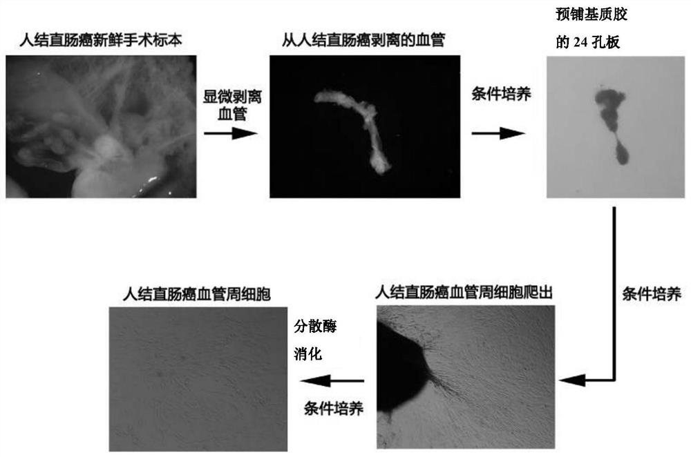
[0072] Example 1 Dissection of human colorectal cancer blood vessels and isolation and culture of human colorectal cancer perivascular cells
[0073] Experimental method: refer to figure 1The flow shown is to take fresh human colorectal cancer samples (collected by the Department of Gastrointestinal Surgery of Guangzhou Overseas Chinese Hospital, and the collected samples were informed of the research purpose and signed the informed consent. According to clinical imaging, serum oncogenic protein test, For malignant colorectal cancer diagnosed preoperatively by biopsy and other technical means, fresh tumor specimens including cancer focus and paracancerous tissue are obtained for surgical resection, and washed with DMEM medium containing 1% (v / v) green chain double antibody to remove residual Pollutants such as feces and blood stains are stored in DMEM medium containing 1% (v / v) penicillin-streptomycin), and in a clean bench, use 1% (v / v) PS (penicillin-streptomycin, penicilli...
Embodiment 2
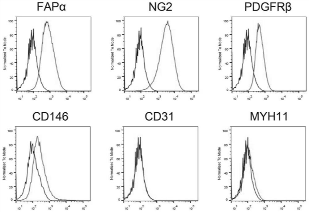
[0075] Example 2 Determination of expression of molecular markers of human colorectal cancer pericytes by flow cytometry
[0076] Experimental method: the human colorectal cancer vascular cells obtained in Example 1 were resuspended with 100 μL of cell staining buffer (staining buffer) and then transferred to a 1.5 mL EP tube, and then 1 μL of anti-CD32-PEblocking (Miltenyi Cat.No.130-097-521) prototype control solution was blocked on ice for 10 minutes; then added anti-FAPα-PE (R&D Cat.No.FAB3715P), anti-NG2-PE (Miltenyi Cat.No.130-097-458), anti-PDGFRβ-PE (Miltenyi Cat.No.130-105-323) and anti-CD146-PE (Miltenyi Cat.No.130-097-939) four pericyte positive molecular markers and anti-CD31-PE (Miltenyi Cat.No.130-110-807), MYH11( Cat.No.PA5-82526) two negative molecular marker flow antibodies, incubate on ice in the dark for 30-60 minutes, wash twice with PBS; add 1 μg / mL DAPI for nuclear staining, keep in the dark on ice Incubate for 10 minutes, wash twice with PBS;...
Embodiment 3
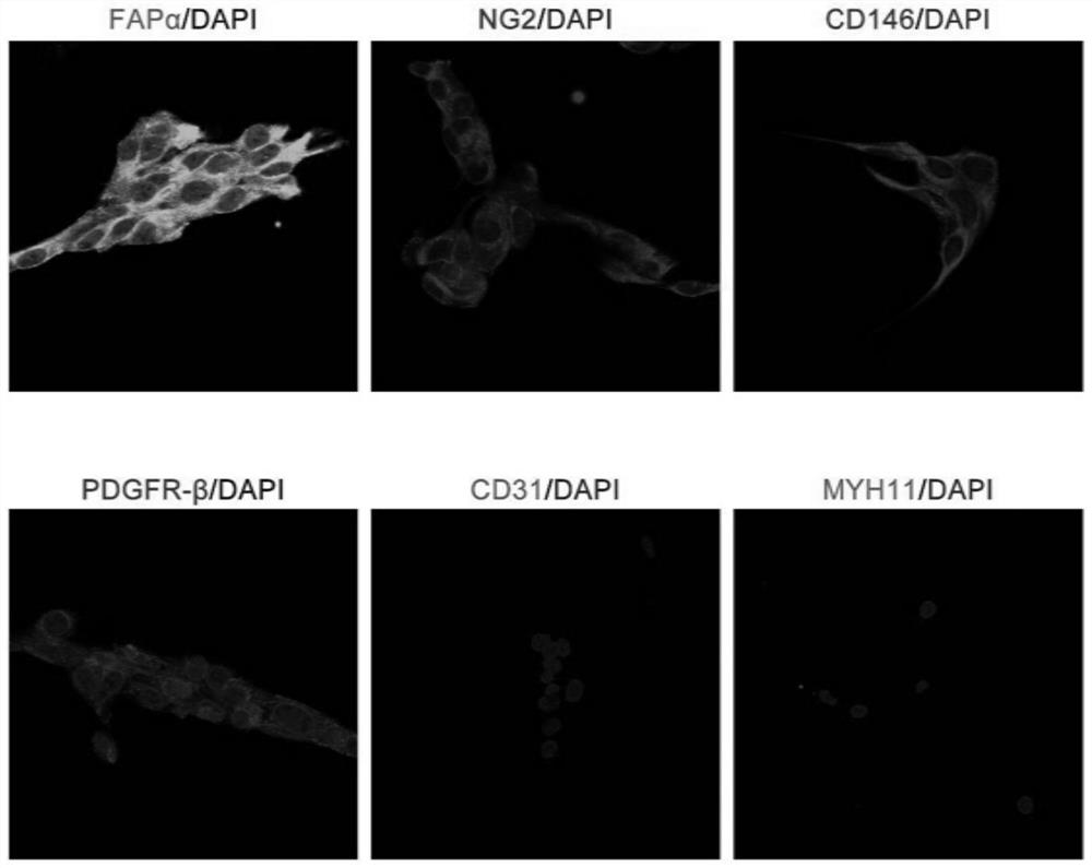
[0078] Example 3 Immunofluorescence assay for the expression of molecular markers of human colorectal cancer perivascular cells
[0079] Experimental method: After the human colorectal cancer perivascular cells obtained in Example 1 were resuspended, 1×10 5 Cells were seeded on laser confocal small dishes at a density of 24 hours, discarded the culture medium and washed with PBS, fixed with 4% (w / v) paraformaldehyde (PBS with a solvent of 0.1M, pH=7.4) at room temperature for 30 minutes, and 0.1 Permeabilize the membrane with %Triton-X100 for 3 minutes, block with 5% BSA at room temperature for 1 hour, add anti-FAPα (R&D Cat.No.AF3715), anti-NG2 (R&D Cat.No.MAB2585), anti-PDGFRβ (R&D Cat.No.AF385) and anti-CD146 (R&D Cat.No.AF932) four pericyte positive molecular markers and anti-CD31 (R&D Cat.No.BBA7), anti-MYH11 ( Cat.No.PA5-82526) the primary antibodies of two negative molecular markers were incubated overnight at 4°C. Wash 3 times with PBS every other day, ea...
PUM
| Property | Measurement | Unit |
|---|---|---|
| Diameter | aaaaa | aaaaa |
Abstract
Description
Claims
Application Information
 - R&D
- Intellectual Property
- Life Sciences
- Materials
- Tech Scout
- Unparalleled Data Quality
- Higher Quality Content
- 60% Fewer Hallucinations
Browse by: Latest US Patents, China's latest patents, Technical Efficacy Thesaurus, Application Domain, Technology Topic, Popular Technical Reports.
© 2025 PatSnap. All rights reserved.Legal|Privacy policy|Modern Slavery Act Transparency Statement|Sitemap|About US| Contact US: help@patsnap.com


