Application of honeycomb-shaped GelMA microspheres in construction of tumor model
A honeycomb and microsphere technology, applied in the field of biomedicine, can solve the problems of lack of molecular markers in the early diagnosis and prognosis of osteosarcoma, the inability to screen drugs in a repeatable and reliable way, and the increase in tumor drug resistance, etc., to achieve maintenance Biological characteristics and gene stability, saving time cost and economic cost, effect of increasing drug resistance
- Summary
- Abstract
- Description
- Claims
- Application Information
AI Technical Summary
Problems solved by technology
Method used
Image
Examples
Embodiment 1
[0078] The preparation of embodiment 1 honeycomb GelMA microsphere
[0079] Experimental materials: Gelatin was purchased from Rousselot (Zhejiang) Co., Ltd. Methacrylic anhydride was purchased from Aladdin Reagents (Shanghai) Co., Ltd.
[0080] A method for preparing honeycomb-shaped GelMA microspheres for constructing a tumor model, the specific steps are as follows:
[0081] (1) Preparation of GelMA: Dissolve 40.0g of gelatin in 400mL of PBS (50°C), accelerate the dissolution on a magnetic stirrer, add 32.0mL of methacrylic anhydride, and pump it in with a micro-syringe pump while stirring, It is used to mediate the displacement reaction between methacryl groups and amino groups and hydroxyl groups on active amino acid residues on gelatin, and then diluted 5 times with 1600 mL of PBS to terminate the reaction. Dialyze for 2 weeks using a dialysis bag with a molecular weight cut-off of 10 kDa, changing ddH every day 2 O, to completely remove free methacrylic anhydride unt...
Embodiment 2
[0084] Example 2 Exploration of the Characterization of Honeycomb GelMA Microspheres
[0085] The microspheres were observed in general by an inverted fluorescence microscope (bright field) and the particle size distribution of the microspheres was calculated by ImageJ; the freeze-dried microspheres were sprayed with gold by ion sputtering, and then observed under SEM and analyzed by ImageJ Calculate the pore size distribution; observe the surface roughness of the honeycomb GelMA microspheres by AFM.
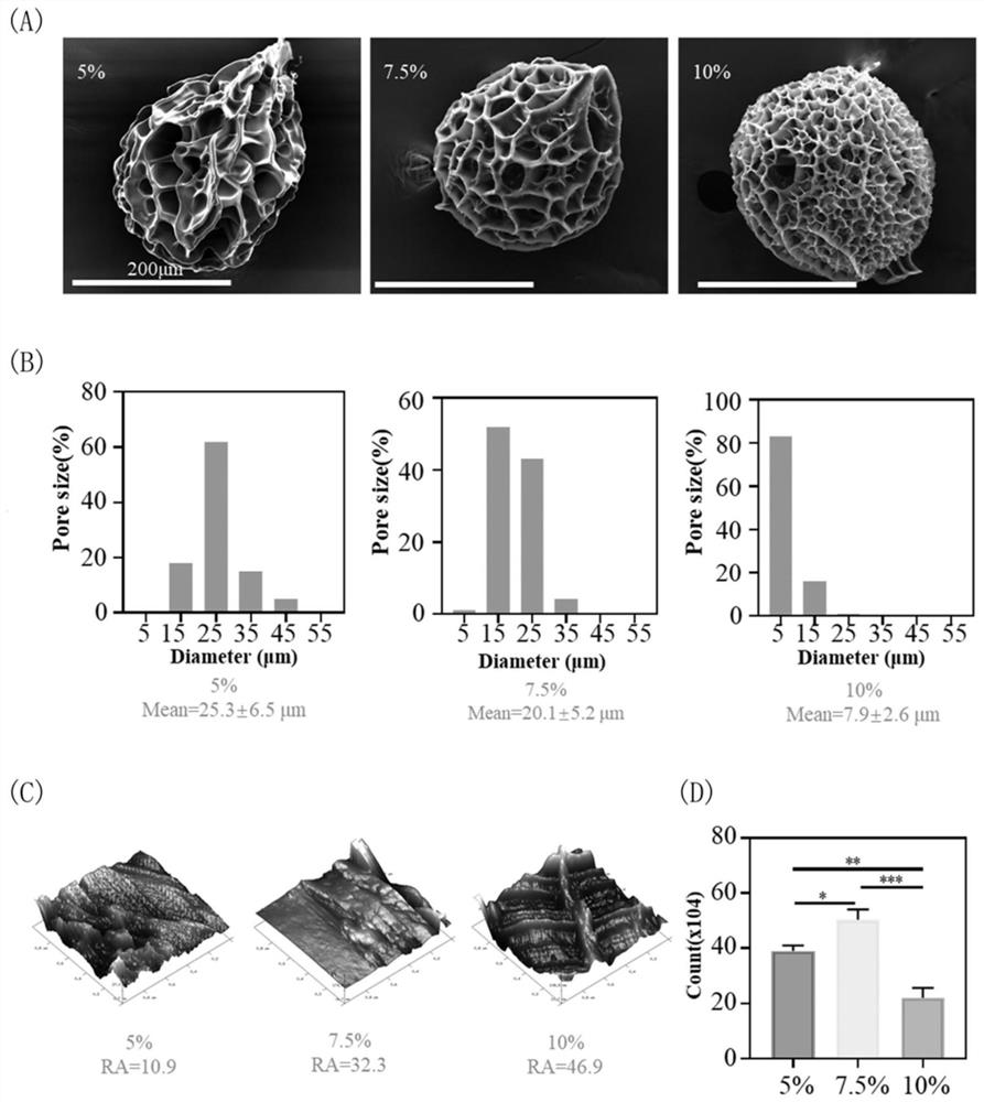
[0086] 1. Under the scanning electron microscope (SEM), the microstructure of microspheres with GelMA concentration of 5%, 7.5% and 10% were observed respectively, and the diameters were all about 350 μm. However, the microspheres produced by different concentrations of GelMA formed different sizes Pore diameter, the average pore diameter of 5%, 7.5% and 10% microspheres is 25.3±6.5μm, 20.1±5.2μm and 7.9±2.6μm, respectively (supporting information such as figure 1 (A) SEM ima...
Embodiment 3
[0088] Example 3 Further Exploration of the Characterization of Honeycomb GelMA Microspheres
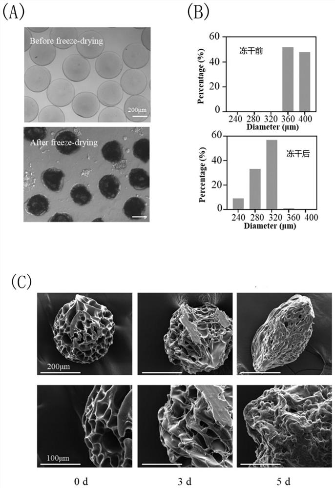
[0089] 1. Use an optical microscope to observe the general view of the microspheres before and after freeze-drying. It can be seen that the microspheres before and after freeze-drying are uniform in size and regular in shape. After contact with water, it will return to the diameter before freeze-drying. This swelling phenomenon provides conditions for the adsorption of cells through swelling during subsequent culture of cells (supporting information such as figure 2 (A), figure 2 (B) shown).
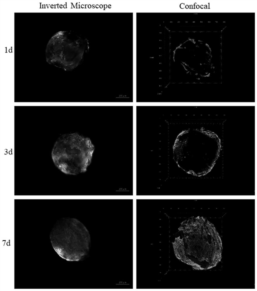
[0090] 2. The microspheres cultured K7M2 cells for 0, 3, and 5 days were dehydrated by ethanol gradient dehydration, and observed under SEM. It can be seen that the microspheres have many pores and are evenly distributed, and the inside and outside are connected, and the cells grow well on the surface of the microspheres And in the pores, the honeycomb-like structure provides more space for...
PUM
 Abstract
Description
Claims
Application Information
 - R&D
- Intellectual Property
- Life Sciences
- Materials
- Tech Scout
- Unparalleled Data Quality
- Higher Quality Content
- 60% Fewer Hallucinations
Browse by: Latest US Patents, China's latest patents, Technical Efficacy Thesaurus, Application Domain, Technology Topic, Popular Technical Reports.
© 2025 PatSnap. All rights reserved.Legal|Privacy policy|Modern Slavery Act Transparency Statement|Sitemap|About US| Contact US: help@patsnap.com


