Hypoxia response co-assembly system based on extracellular vesicles and preparation method thereof

A technology of co-assembly and vesicles, applied in the biological field, can solve the problems of inconvenient use, body damage, complex space for special equipment, etc.

Active Publication Date: 2021-12-24
NANKAI UNIV
View PDF5 Cites 0 Cited by
  • Summary
  • Abstract
  • Description
  • Claims
  • Application Information

AI Technical Summary

Problems solved by technology

However, excessive hypoxic signals can lead to body damage and even death
In view of the deepening understanding of hypoxia on diseases, researchers are paying more and more attention to the clinical application of tracer tissue hypoxia, such

Method used


Image

  • Hypoxia response co-assembly system based on extracellular vesicles and preparation method thereof
  • Hypoxia response co-assembly system based on extracellular vesicles and preparation method thereof
  • Hypoxia response co-assembly system based on extracellular vesicles and preparation method thereof

Examples


Embodiment 1

[0075] This example is used to illustrate a method for extracting extracellular vesicles derived from umbilical cord-derived mesenchymal stem cells.

[0076] The processing steps are as follows: place the fetal bovine serum in an ultracentrifuge tube, centrifuge at 100,000 g at 4°C for 2 hours, take the supernatant in an ultra-clean bench, filter it with a 0.22 μm needle filter, and store it in a -80°C refrigerator for later use.

[0077] See "Animal Cell Culture (Sixth Edition)" for details on the operation steps of cell biology experiments such as cell subculture, cryopreservation and recovery.

[0078] Collect the conditioned medium of human umbilical cord-derived mesenchymal stem cells (hP-MSCs) containing extracellular vesicles: when cultured in 75cm 2 The hP-MSCs in the cell culture flask are in the logarithmic growth phase. When the confluence of the cells reaches 80%, the medium is aspirated, washed twice with PBS, and 10ml of prepared 10% extracellular vesicle-free F...

Embodiment 2

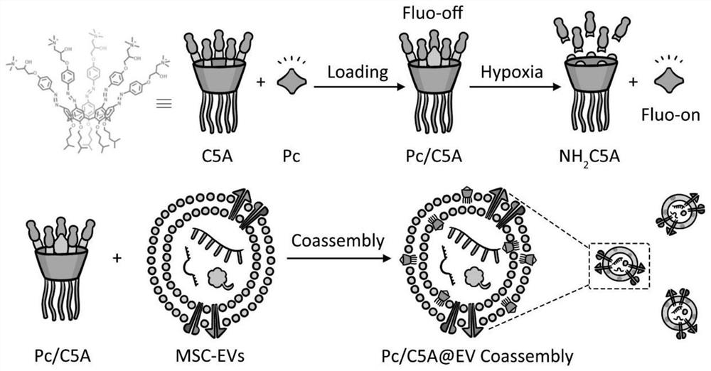
[0085] This example is used to illustrate a method for preparing a co-assembly system based on extracellular vesicles (EVs) for hypoxia response imaging.

[0086] (1) After mixing aluminum phthalocyanine Pc and calixarene C5A respectively, incubate in a dark room at 37°C for 30 minutes.

[0087] (2) Add Pc / C5A to the extracted 100 μL EVs sample containing 200-300 μg protein, and make up to 500 μL with PBS. At this time, the final concentration of Pc / C5A is 10 μM / 20 μM. Educate for 2h.

[0088] (3) Transfer the above mixture into an ultracentrifuge tube, fill it up with PBS, centrifuge at 100,000 g at 4°C for 120 minutes, remove the supernatant, and obtain extracellular vesicles that have been stained green.

[0089] (4) Extracellular vesicles were resuspended in 50 μl PBS, aliquoted and stored in a -80°C refrigerator for later use.

Embodiment 3

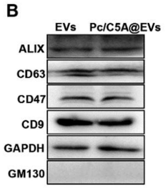
[0091] This example is used to illustrate an identification method of a hypoxia response imaging co-assembly system based on extracellular vesicles (EVs).

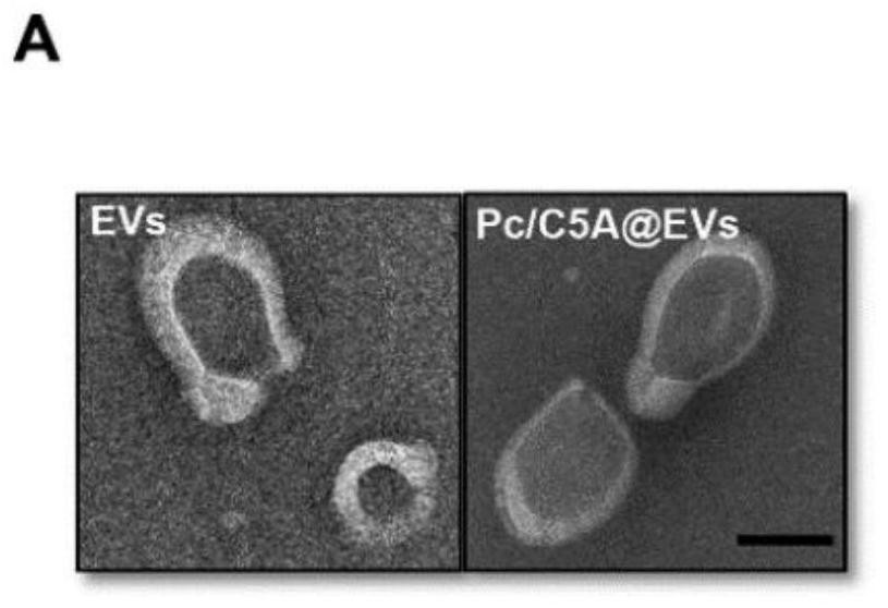
[0092] (1) Identification of extracellular vesicle morphology using transmission electron microscopy

[0093] The extracellular vesicles extracted in Example 1 and the hypoxia-response imaging co-assembly system in Example 2 were respectively dropped on a 200-mesh sample copper grid, left at room temperature for 2 minutes, and the excess liquid was blotted with filter paper; 20 mg / mL was added dropwise on the sample grid Uranium acetate solution, let stand at room temperature for 1 min, negatively stain the sample, blot the excess liquid with filter paper, and dry the sample net; observe the prepared sample under a transmission electron microscope, and collect photos. Such as figure 1 As shown, the shape and diameter of extracellular vesicles did not change, and the shape was a cup-shaped vesicle-like structure with a dia...


PUM

PropertyMeasurementUnit
Diameteraaaaaaaaaa

Abstract

The invention relates to a co-assembly system based on extracellular vesicles and used for hypoxia response imaging. The co-assembly system comprises the extracellular vesicles and Pc/C5A attached to the extracellular vesicles, and the Pc/C5A contains calixarene QAC5A-6C and aluminum phthalocyanine AlPcS4. The method comprises the following steps: mixing calixarene QAC5A-6C (C5A) and aluminum phthalocyanine AlPcS4 (Pc), and carrying out first incubation to obtain Pc/C5A; and mixing with extracellular vesicles, and carrying out second incubation, so as to obtain the co-assembly system for hypoxia response imaging.

Description

technical field [0001] The present disclosure relates to the field of biotechnology, in particular, to an extracellular vesicle-based co-assembly system for hypoxia response imaging, and a method for preparing an extracellular vesicle-based co-assembly system for hypoxia response imaging Methods and a self-assembled Pc / C5A based on co-assembly with extracellular vesicles aimed at imaging the hypoxia response. Background technique [0002] Oxygen is an essential element in the energy metabolism process of all living organisms. Many physiological processes are regulated by oxygen, which makes the body's perception and regulation of changes in oxygen concentration in the environment very precise. Hypoxia is the key cause of many diseases, such as tumors, anemia, cardiovascular and cerebrovascular diseases, etc. In fact, mild hypoxia mainly causes compensatory responses of living organisms, and the hypoxic signaling pathway has a protective effect on some tissue oxidative damag...

Claims


Application Information

Patent Timeline
IPC IPC(8): C09K11/06C09K11/02A61K49/00G01N21/64
CPCC09K11/06C09K11/025A61K49/0036A61K49/0052A61K49/005G01N21/6428C09K2211/1074C09K2211/186
Inventor 王悦冰郭东升程元秋岳宇昕王斓星
Owner NANKAI UNIV
Why Eureka
Try Eureka