Cell nucleus fluorescent dye and dyeing method thereof

A technology of fluorescent dyes and dyeing methods, which is applied in the direction of luminescent materials, organic dyes, fluorescence/phosphorescence, etc., can solve the problems of inconvenient operation and complicated dyeing procedures, and achieve easy operation, good photostability, and simple dyeing procedures. Effect

Pending Publication Date: 2022-02-18
SHENZHEN INST OF ADVANCED TECH
View PDF2 Cites 2 Cited by
  • Summary
  • Abstract
  • Description
  • Claims
  • Application Information

AI Technical Summary

Problems solved by technology

Carefully analyze the nuclear fluorescent dyes commonly used in the domestic and foreign markets at present, and there are not many compound systems that meet the above requirements, and some dyeing procedures are complicated, need to be washed, and are not easy to operate

Method used

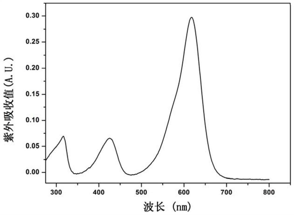
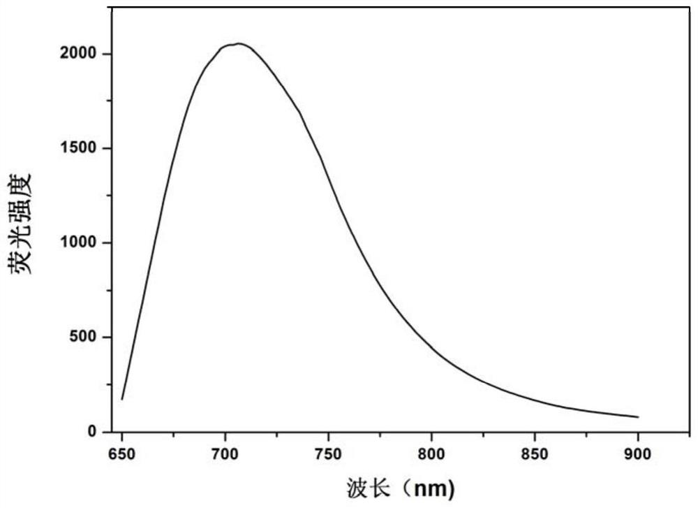
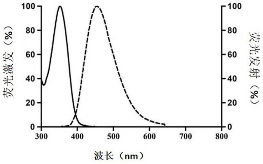

Image

  • Cell nucleus fluorescent dye and dyeing method thereof
  • Cell nucleus fluorescent dye and dyeing method thereof
  • Cell nucleus fluorescent dye and dyeing method thereof

Examples


Embodiment 1

[0073] Example 1 For staining of fixed cells or tissues

[0074] The dyeing method is:

[0075] Prepare a PBS buffer solution of the nuclear fluorescent dye of the present invention with a stock solution of 1 mg / mL.

[0076] a. Cell or tissue samples, after fixation, are properly washed to remove the fixative. Add the staining solution of the nuclear fluorescent dye of the present invention at a concentration of 100 μg / mL to cover the sample. Place at room temperature for 3-5min. No need to wash, observe directly under a fluorescent microscope or under a fluorescent confocal microscope after mounting.

[0077] b. For adherent cells or tissue sections, add the staining solution of the nuclear fluorescent dye of the present invention at a concentration of 100 μg / mL to cover the sample. For suspension cells, add staining solution at least 3 times the volume of the sample to be stained, and mix well. Place at room temperature for 3-5min. No need to wash, observe directly und...

Embodiment 2

[0078] Example 2 For staining of living cells or cultured tissues

[0079] a. Add the staining solution of the nuclear fluorescent dye of the present invention with a concentration of 100 μg / mL, which must fully cover the sample to be stained. Usually, 1 mL of staining solution needs to be added to one well of a 6-well plate, and 100 μL needs to be added to one well of a 96-well plate. staining solution.

[0080] b. Incubate at a temperature suitable for cell culture for 5 minutes, then perform fluorescence detection.


PUM

PropertyMeasurementUnit
concentrationaaaaaaaaaa
wavelengthaaaaaaaaaa
concentrationaaaaaaaaaa

Abstract

The invention provides a cell nucleus fluorescent dye as well as a dyeing method and application thereof, and provides a novel fluorescent dye for cell nucleus dyeing. The cell nucleus fluorescent dye has good membrane permeability, high sensitivity, rapid nucleus dyeing efficiency, good light stability, good biocompatibility and longer-wavelength exciting light and emission light, and can be applied to fluorescent dyeing of cell nucleuses.

Description

technical field [0001] The invention relates to the technical field of fluorescence imaging, in particular to a cell nucleus fluorescent dye and a staining method thereof. Background technique [0002] Bioimaging generally refers to methods for visualizing and recording target molecules or processes in cells, tissues, or in vivo. Bioimaging technology includes fluorescence imaging technology, Raman imaging technology, nuclear magnetic resonance technology, photoacoustic imaging technology, ultrasonic imaging technology, X-ray photography technology and positron emission tomography technology, etc. Among them, fluorescence imaging techniques including confocal microscopy, two-photon microscopy, and super-resolution microscopy have won extensive research interest due to their low damage to samples, excellent sensitivity, and high spatial-temporal resolution. In bioimaging, for different biomolecules and biological processes, many luminescent bioprobes have been designed and a...

Claims


Application Information

Patent Timeline
Patent Type & Authority Applications(China)
IPC IPC(8): C07C251/20C09K11/06C09B11/12G01N1/30G01N21/64
CPCC07C251/20C09K11/06C09B11/12G01N21/6428G01N1/30C09K2211/1007G01N2021/6439G01N2001/302
Inventor 龚萍周理华张鹏飞蔡林涛
Owner SHENZHEN INST OF ADVANCED TECH
Why Eureka
Try Eureka