A fully human monoclonal antibody against novel coronavirus and its application
A monoclonal antibody and coronavirus technology, applied in antiviral agents, antiviral immunoglobulins, antibodies, etc., can solve the problems of limited plasma sources of recovered patients, the risk of disease infection due to batch-to-batch differences, and the inability to popularize and apply it to achieve inhibition Effect of virus infecting host cells, good druggability, high and high activity
- Summary
- Abstract
- Description
- Claims
- Application Information
AI Technical Summary
Problems solved by technology
Method used
Image
Examples
Embodiment 1
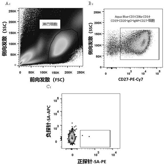
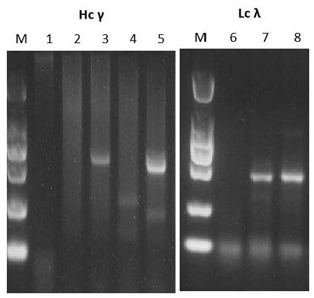
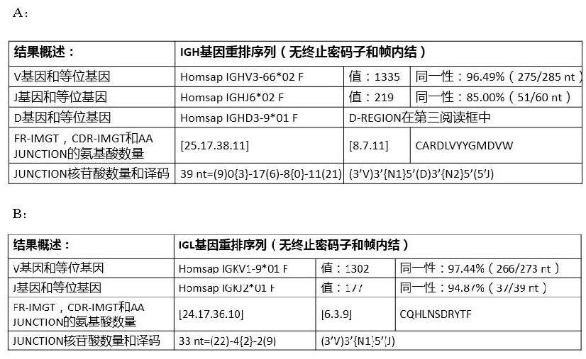
[0064] Example 1: Screening and preparation of human anti-SARS-CoV2 monoclonal antibodies
[0065] 1.1 Labeling of probes
[0066] The required SARS-CoV2-related specific positive and negative probes are conjugated with fluorescent dyes, and the process is as follows:
[0067] 1.1.1 Use AviTag Fusion Protein Biotinylation Kit (Avidity, BirA500) for biotin labeling of the probe. The reaction system was prepared according to the procedure described in the manual and incubated at 30 °C for 30 min.
[0068] 1.1.2 Use a 30 kDa ultrafiltration tube to exchange the reaction product with 1× PBS (pH 7.4) to remove excess biotin.
[0069] 1.1.3 Gradually add 1 / 5 molar equivalent of streptavidin-phycoerythrin (SA-PE) or streptavidin-allophycocyanin (SA-APC) to the biotin label at 20 min intervals Among the probes, until the molar ratio of SA-PE or SA-APC to biotin-labeled probe reaches 1:1, incubate at 4 °C with gentle shaking to prepare positive and negative probes labeled with fluor...
Embodiment 2
[0100] Example 2: Analysis of the binding activity of monoclonal antibody YK-CoV2-03 to RBD protein
[0101] Antibody and Antigen Binding Activity Analysis Using Gator Unlabeled Biomolecule Analyzer
[0102] 2.1. Using GNTi cells to express RBD, after purification by nickel column and molecular sieve in turn, biotinylated RBD according to the steps shown in the instructions of DSB-X™ Biotin Labeling Kit (Invitrogen™, D20655).
[0103] 2.2 Dilute the biotinylated RBD by 600 times, add 3 μL of RBD to 2000 μL of HBS-EP buffer and mix, and load the diluted RBD into a 96-well sample plate in an amount of 200 μL per well.
[0104] 2.3 Use HRV 3C protease to cut the monoclonal antibody into Fab fragments, use affinity chromatography and molecular sieve to purify the obtained antibody Fab, after diluting the antibody Fab 1000 times, continue to carry out gradient dilution according to 1:3, a total of 4 dilutions (36.2nM, 12.1nM, 4.02nM and 1.34nM), take 200 μL of each dilution sample...
Embodiment 3
[0109] Example 3: Analysis of the ability of monoclonal antibody YK-CoV2-03 to competitively block the binding of SARS-CoV2 virus RBD to receptor ACE2
[0110] Analysis of the binding ability of antibodies to block SARS-CoV2 virus RBD and receptor ACE2 using Gator non-labeled biomolecule analyzer
[0111] 3.1 Expi-CHO cells were used to express the receptor ACE2, and after purification by nickel column and molecular sieve in sequence, ACE2 was biotinylated according to the steps shown in the instructions of DSB-X™ Biotin Labeling Kit (Invitrogen™, D20655). Biotinylated ACE2 was diluted to 40 μg / mL at a molar concentration of 570 nM.
[0112] 3.2 Dilute RBD to 30 μg / mL with a molar concentration of 1000 nM, dilute the antibody to 0.2 mg / mL, and mix with RBD in a molar ratio of 1:1. The molar amount of antibody in the prepared mixture is greater than the molar amount of ACE2. .
[0113] 3.3 During the detection process, each experiment was first equilibrated in the HBS-EP buff...
PUM
 Abstract
Description
Claims
Application Information
 - R&D
- Intellectual Property
- Life Sciences
- Materials
- Tech Scout
- Unparalleled Data Quality
- Higher Quality Content
- 60% Fewer Hallucinations
Browse by: Latest US Patents, China's latest patents, Technical Efficacy Thesaurus, Application Domain, Technology Topic, Popular Technical Reports.
© 2025 PatSnap. All rights reserved.Legal|Privacy policy|Modern Slavery Act Transparency Statement|Sitemap|About US| Contact US: help@patsnap.com


