

Method of separating pork liver stem cell
A stem cell and pig liver technology, applied in the field of pig liver stem cells, can solve the problems of low cell purity, small number of cells, long time required, etc., and achieve the effect of good survival rate and high purity
- Summary
- Abstract
- Description
- Claims
- Application Information
AI Technical Summary
Problems solved by technology
Method used
Image

Examples
Embodiment 1
[0036] Embodiment 1, using the method of the present invention to isolate porcine liver stem cells
[0037] Experimental pigs underwent partial liver resection to promote liver regeneration. The steps are as follows:
[0038] 1) Use 20 mg / kg of pentobarbital sodium per kilogram of test pig body weight to carry out abdominal anesthesia to test pigs;
[0039] 2) Autoclave the surgical instruments used, and ultraviolet disinfection in the operating room;
[0040] 3) Cut off the hair on the abdominal cavity body surface of the test pig with electric clippers, disinfect the body surface of the test pig with 70wt% alcohol, and spread a towel;
[0041] 4) Cut the epidermis, and then dissect the abdominal cavity. Be careful not to cut the pleural membrane, so as not to cause death due to pneumothorax;
[0042] 5) Fix the epidermis and abdominal muscles with hemostats, add 0.90wt% normal saline dropwise into the abdominal cavity, push the mesentery to the left, add 0.90wt% normal salin...
Embodiment 2
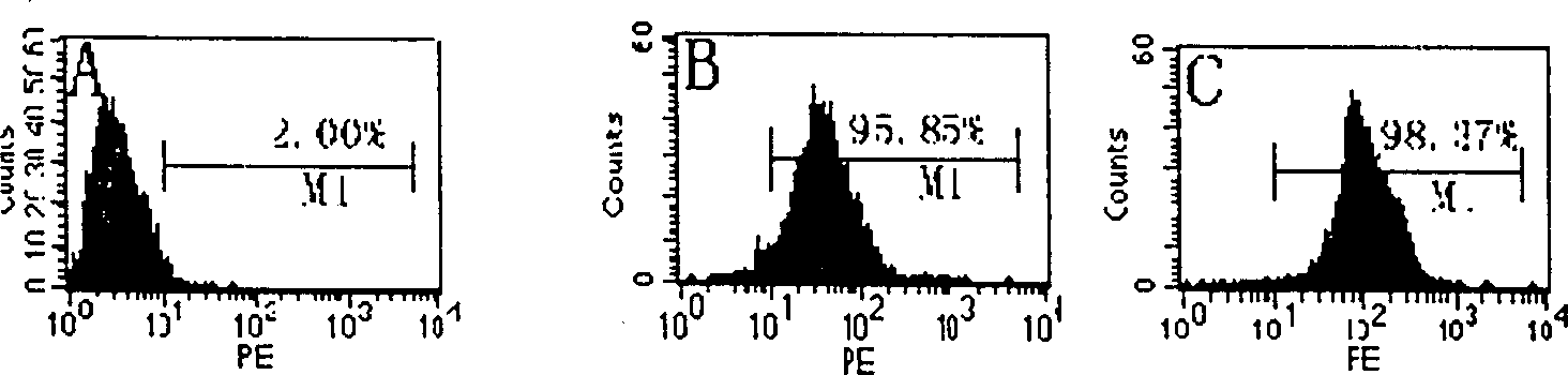
[0061] Embodiment 2, identifying and analyzing porcine liver stem cells with flow cytometry (single staining detection)
[0062] Hepatic stem cells from 2 pigs were isolated according to the method in Example 1.
[0063] The porcine liver stem cells were identified by flow cytometry: freshly separated cells were added to eppendoff tubes (10 5 cells / tube), centrifuge at 1500rpm for 10min, wash once with PBS; loosen the cell pellet, add 1ml of cold acetone, fix at 4°C for 8min; centrifuge at 1500rpm for 5min, wash with PBS three times, and centrifuge at 1500rpm for 10min; Add 0.1ml PBS to make a cell suspension, add primary antibody albumin and CK-19 dropwise, and incubate at 37°C for 45 minutes; centrifuge at 1500rpm for 10 minutes, discard the supernatant, add 0.1ml PBS to the precipitate to make a cell suspension; Add PE-labeled IgG dropwise, incubate at 37°C for 30 minutes, wash with PBS 3 times; centrifuge at 1500rpm for 10 minutes, add 0.5-1ml PBS to the sediment dependin...
Embodiment 3
[0065] Embodiment 3, identifying and analyzing porcine liver stem cells with flow cytometry (double staining detection)
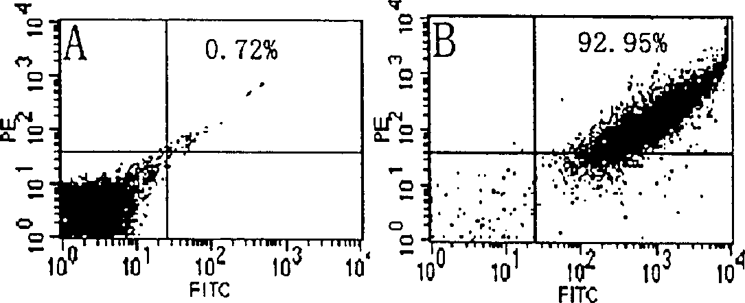
[0066] According to the method in Example 1, 6 days after partial hepatectomy, pig liver stem cells were isolated from 3 test pigs according to the method in Example 1, and each test pig obtained an average of 5.3 × 10 7 cells.
[0067] The double staining procedure is similar to the single staining in Example 2, except that the primary antibody is albumin + CK-19, and the secondary antibody includes FITC-labeled rabbit anti-mouse IgG (binding to CK-19) and PE-labeled goat antibody Rabbit IgG (albumin bound). Negative controls were replaced with FITC-labeled rabbit anti-mouse IgG and PE-labeled goat anti-rabbit IgG.
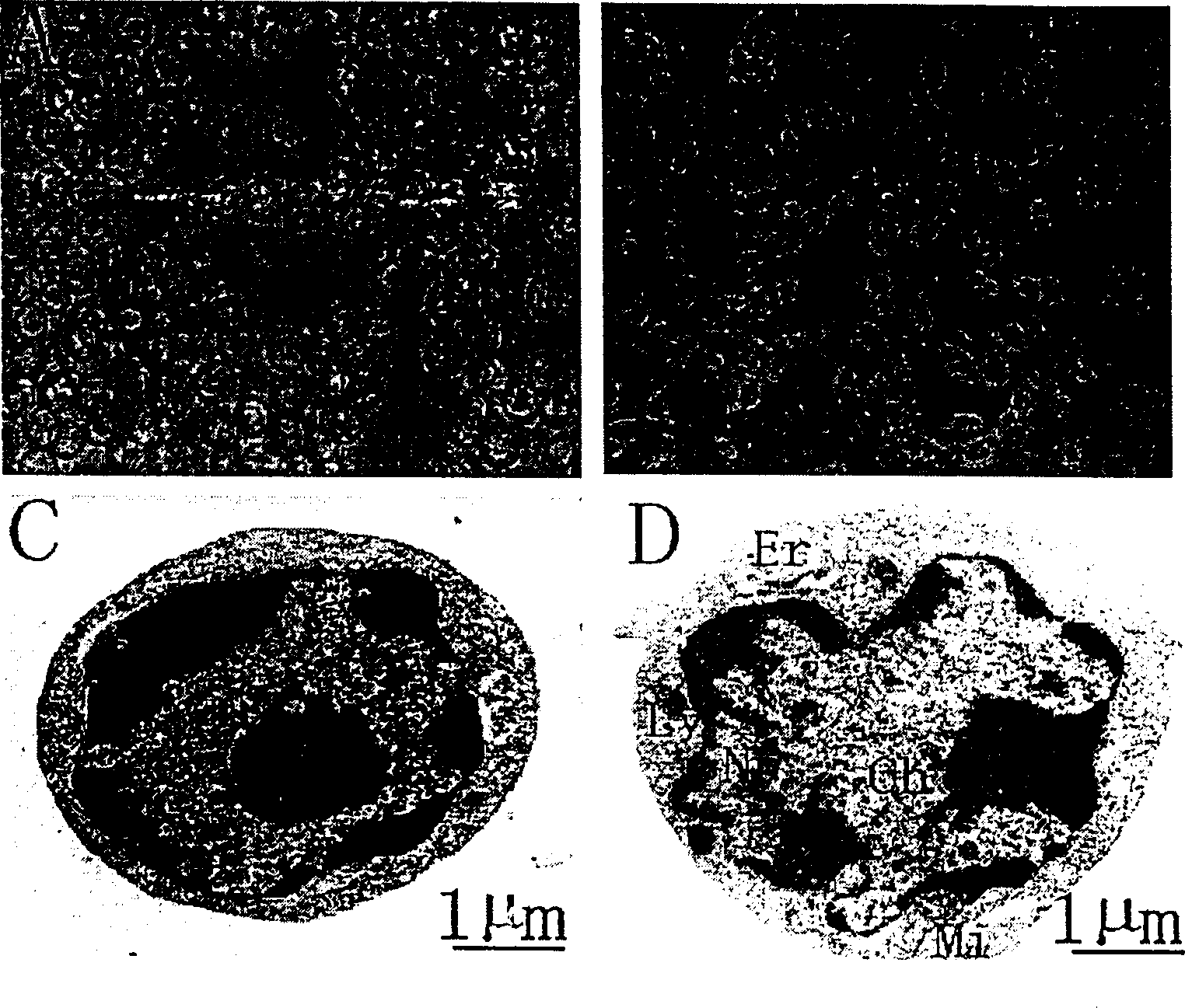
[0068] Biochemical phenotypes of freshly isolated porcine liver stem cells analyzed by flow cytometry, plotted in image 3 . The results of double staining showed that 92.23% of the cells co-expressed albumin and CK-19 (such as image 3 B...
PUM
| Property | Measurement | Unit |
|---|---|---|
| diameter | aaaaa | aaaaa |
Abstract
Description
Claims
Application Information