

Double-layered artificial skin and its prepn process
A technology of artificial skin and dermis, applied in the field of medicine and biomedical engineering, can solve the problems of difficult operation, large shrinkage rate of collagen gel, unpredictable immune rejection, etc.
- Summary
- Abstract
- Description
- Claims
- Application Information
AI Technical Summary
Problems solved by technology
Method used
Image

Examples
Embodiment 1
[0058] Isolation and culture of keratinocytes
[0059] 1) Take materials
[0060] The surgically removed foreskin of children is placed in the keratinocyte culture medium containing antibiotics and brought into the cell culture room.
[0061] 2) Separate the epidermis
[0062] Carefully cut off the subcutaneous fat tissue and part of the dermis with scissors, and trim them into strips with a width of about 1-2mm, rinse twice with calcium and magnesium-free PBS, and soak in 0.24U / ml neutral protease (Dispase II) with the epidermis facing upward overnight at 4°C.
[0063] 3) digestion
[0064] After 18 hours, the epidermis was torn off from the dermis with two ophthalmic tweezers, cut into pieces, and then digested with 0.05% trypsin-0.53mM EDTA at 37°C for 15 minutes, and 10ml trypsin inhibitor (10mg / ml) to stop the digestion . Filter through a 150-mesh stainless steel filter, centrifuge at 1000r / min for 10 minutes, discard the supernatant, add 10ml of keratinocyte culture...
Embodiment 2
[0070] Isolation and culture of dermal fibroblasts
[0071] 1) digestion
[0072] Cut the dermis after peeling off the epidermis into pieces, digest with 0.1% collagenase at 37°C for 2 hours, filter through a 150-mesh stainless steel filter, centrifuge at 1000r / min for 5 minutes, discard the supernatant, and add calcium-free magnesium Wash 3 times with PBS. Add 10ml of DMEM culture solution containing 10% fetal bovine serum, mix well, and trypan blue staining and counting.
[0073] 2) Primary culture
[0074] Make a single cell suspension after counting, press 1-2×10 6 / 75cm 2 Density inoculated culture, DMEM medium containing 10% fetal bovine serum, at 37°C, 5% CO 2 , cultured in an incubator with 100% relative humidity, and the culture medium was changed every 3 days.
[0075] 3) Subculture
[0076] When the cells are confluent to 60%-75% density, wash with 10ml calcium and magnesium-free PBS, digest with 0.25% trypsin at 37°C for 5 minutes, add DMEM culture medium co...
Embodiment 3
[0078] Preparation of artificial skin
[0079] (1) Construction of fibroblast-PGA complex
[0080] The dermal fibroblasts prepared in Example 2 were collected by trypsin digestion, and then were divided into 0.2 × 10 6 / 10mg polyglycolic acid, 1×10 6 / 10mg polyglycolic acid, 5×10 6 It was inoculated on a network of polyglycolic acid (PGA) at a concentration of 10 mg polyglycolic acid. Then inoculate the inoculated PGA material at 37±0.5°C, 5% CO 21. Under the condition of saturated humidity, culture in DMEM culture solution containing 10% fetal bovine serum (the culture solution is changed every 3 days) for 1-2 weeks to form fibroblast-PGA complex.
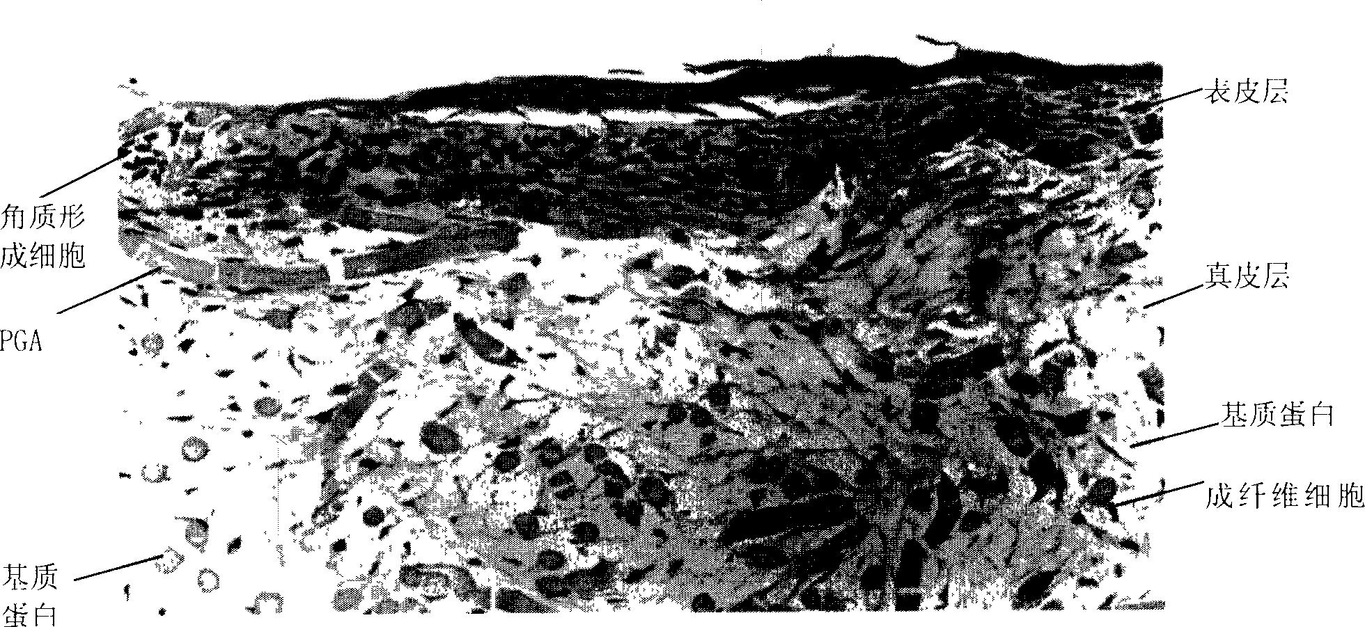
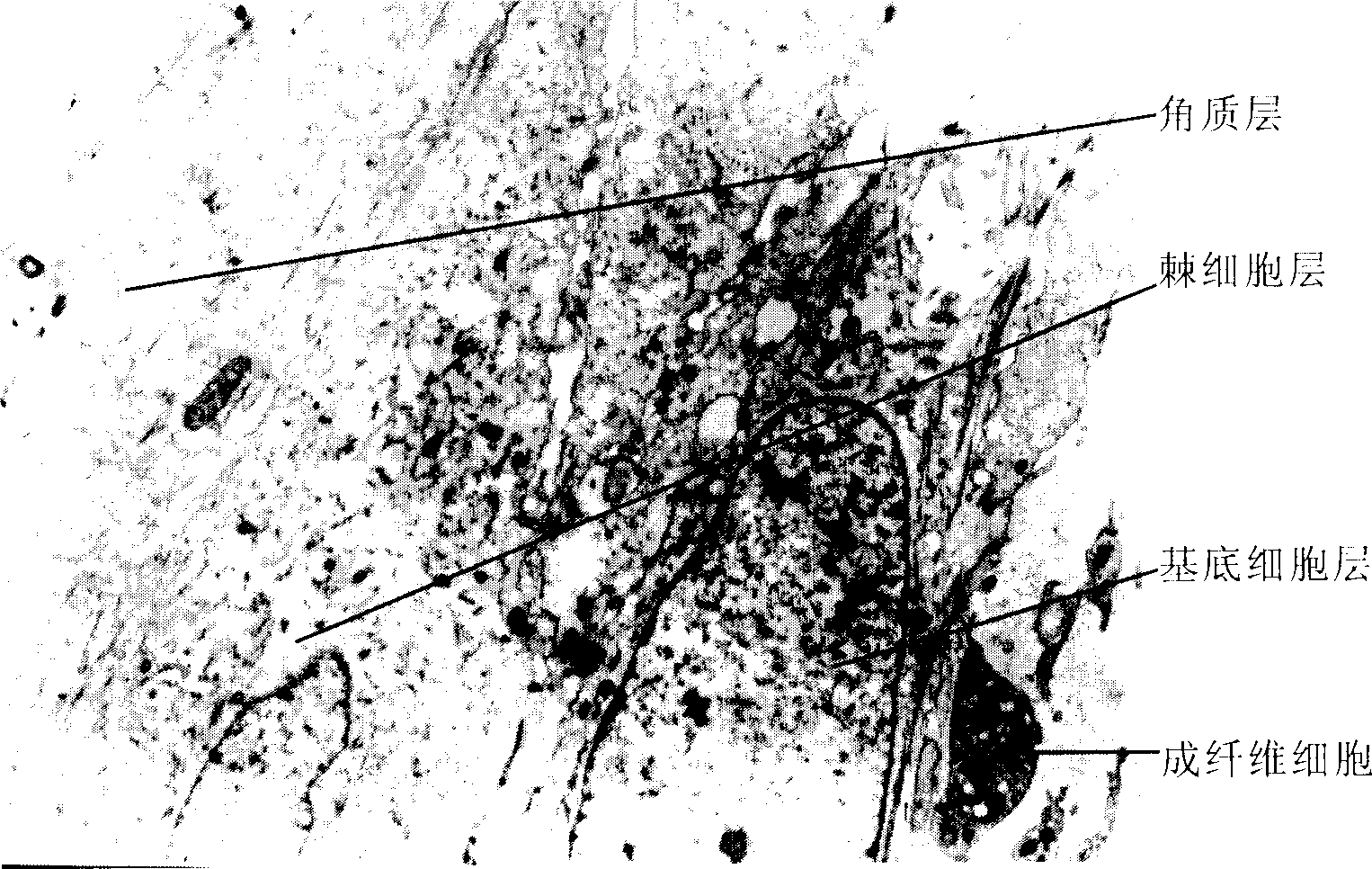
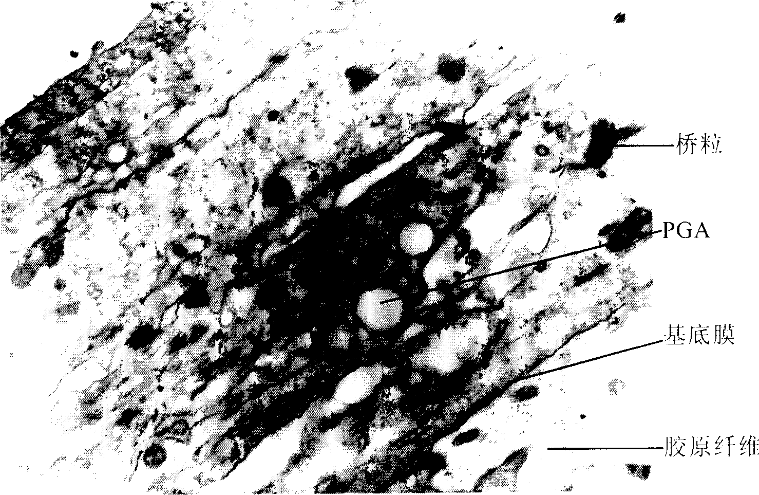
[0081] (2) Formation of double skin
[0082] The keratinocytes prepared in Example 1 were collected, and then respectively divided into 0.5×10 5 / square centimeter, 5×10 5 / square centimeter, 50×10 5 / cm 2 seeded on the surface of the fibroblast-PGA complex prepared in the previous step. The inoculated complex was mixed ...
PUM
| Property | Measurement | Unit |
|---|---|---|
| Thickness | aaaaa | aaaaa |
| Thickness | aaaaa | aaaaa |
Abstract
Description
Claims
Application Information