Muramidase detction reagent and its preparing method

A technology for detecting reagents and lysozyme, applied in the field of biochemical analysis, can solve the problems of increasing the difficulty of operation, difficult to adapt to the development trend of detection requirements, and the influence of subjective errors of test results operators.

Inactive Publication Date: 2005-03-02
SHANGHAI CHEST HOSPITAL
View PDF0 Cites 4 Cited by
  • Summary
  • Abstract
  • Description
  • Claims
  • Application Information

AI Technical Summary

Problems solved by technology

Among them, the standardization of reagents in the plate method is difficult, the operation is cumbersome, and specific training is required to master it, and the test results are easily affected by the subjective error of the operator; the turbidimetric method is all manual operation, and the bacterial suspension used in the manual turbidimetric method is left standing. Afterwards, the bacteria will sink, so the bacter

Method used


Image

  • Muramidase detction reagent and its preparing method
  • Muramidase detction reagent and its preparing method

Examples


Embodiment 1

[0020] Example 1 Preparation of lysozyme test reagent

[0021] 1. Use conventional culture methods to cultivate micrococcus on nutrient agar for 48 hours,

[0022] 2. Under aseptic conditions, wash the lawn with physiological saline,

[0023] 3. Use active chlorine to inactivate, the concentration is 1000ppm, the time is 30 minutes,

[0024] 4. Filter the inactivated micrococcus with gauze to remove the mixed agar fragments; wash the bacteria with normal saline, and then wash with 0.125mol / L pH6.0 phosphate buffer.

[0025] 5. Suspend with a phosphate buffer containing 1.3‰ gelatin and 1‰ sodium azide, and use the same buffer to adjust the bacterial suspension to a light path of 1cm and an absorbance of about 1.5A at a wavelength of 650nm to obtain a bacterial suspension. Store at 4°C to obtain the lysozyme test reagent of the present invention.

Embodiment 2

[0026] Example 2 Preparation of lysozyme test reagent

[0027] Use conventional culture methods to culture micrococcus on nutrient agar for 72 hours, wash the lawn with physiological saline under aseptic conditions, inactivate it with active chlorine at a concentration of 2000ppm for 15 minutes, filter the inactivated micrococcal gauze to remove the mixture Agar fragments; wash the bacteria with physiological saline, then wash with 0.0625mol / L pH6.0 phosphate buffer, suspend with 1.5‰ gelatin, 1‰ sodium azide in phosphate buffer, and use the same buffer Adjust the bacterial suspension to a light path of 1 cm and an absorbance of about 1.5 A at a wavelength of 650 nm to obtain the bacterial suspension. Store at 4°C to obtain the lysozyme test reagent of the present invention.

Embodiment 3

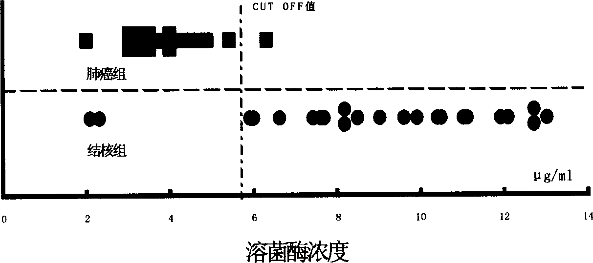
[0028] Example 3 Clinical test

[0029] Fifty adult physical examination serum specimens were taken; 44 cases of pleural effusion specimens were taken. There were 21 cases of cancerous pleural effusion, all confirmed by cytology, 19 cases were lung adenocarcinoma, 1 case was suspected to be adenocarcinoma, 1 case was postoperative breast cancer; 23 cases were tuberculous pleural effusion. After the specimens are collected, they are frozen at -40°C. When testing, place at room temperature to order re-thawing, invert to mix, centrifuge and take the supernatant for inspection.

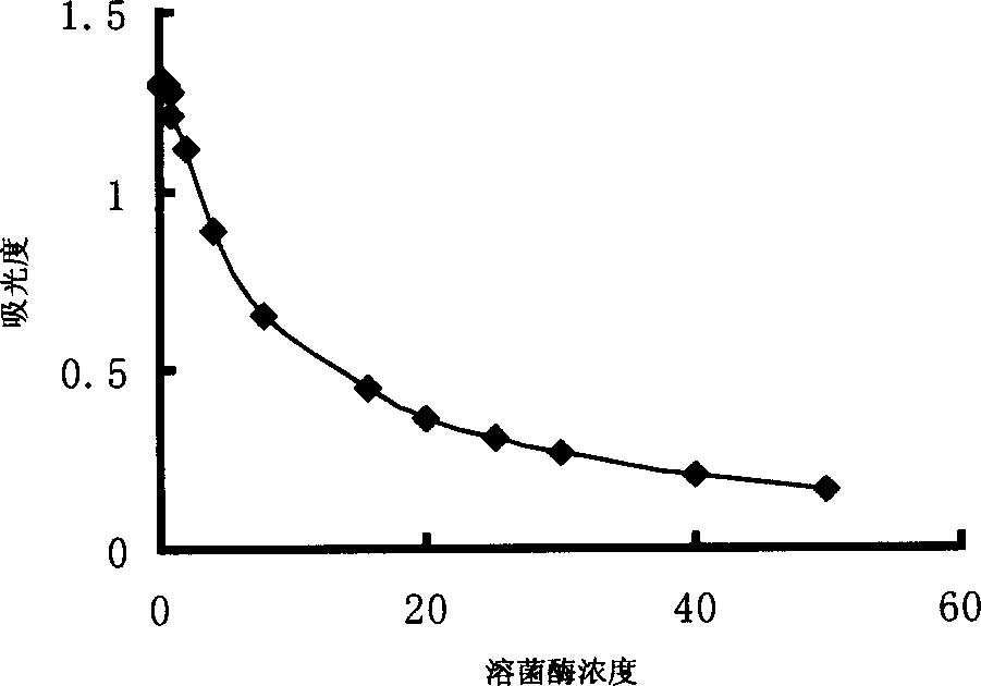
[0030] Use HITACHI 7060 automatic biochemical analyzer to establish an automatic analysis method for lysozyme activity. Method: endpoint method; wavelength: 340 / 623nm; reagent volume: 250μl; sample volume: 30μl; measurement points: 31 points. The lysozyme standard was prepared into a 15.6μg / ml solution as a calibration solution, and a 5-point calibration was performed.

[0031] Dilute the standard lysozyme us...


PUM


Abstract

The invention of biochemical analysis field relates to a lysozyme testing agent and its manufacture method. The lysozyme testing agent contains killing Micrococcus and gelatin, which is bacteria suspension with high stability, created by new method of killing Micrococcus and using gelatin as medium and stabilizer of disperse thallus. After more than four hours of standing, if absorbance of bacteria suspension does not change notably, agent abtained is proved to have excellent stability and can satisfy various testing requirement, be used in automatic biochemical analysis and realize automatic and quantitive detection of lysozyme activity.

Description

Technical field [0001] The invention belongs to the field of biochemical analysis, and specifically relates to a lysozyme detection reagent and a preparation method thereof Background technique [0002] Lysozyme analysis is usually used for the detection of various body fluids. Its significance is to judge the change and severity of the disease through the determination and analysis of lysozyme in body fluids. According to the authoritative book "Methods in Clinical Chemistry" edited by Pesce A.J. and Kaplan L.A. (translated by Lin Qisui and Wen Qingcheng), the main methods currently used for clinical lysozyme analysis are plate method and turbidimetric method. Among them, the standardization of reagents by the plate method is difficult, the operation is cumbersome, and it requires specific training to master, and the test results are easily affected by the subjective error of the operator; the turbidimetric method is all manual operation, and the bacterial suspension used in the...

Claims


Application Information

Patent Timeline
IPC IPC(8): C12Q1/00C12Q1/527G01N21/31
Inventor 于方治吴京
Owner SHANGHAI CHEST HOSPITAL
Why Eureka
Try Eureka