Intervention type tissue hardness supersonic obtaining method and intervening supersonic hardness-detecting device

A technology of tissue hardness and detector, which is applied in ultrasonic/sonic/infrasonic diagnosis, sonic diagnosis, infrasonic diagnosis, etc. It can solve problems such as low accuracy, large size, and inflexibility, and achieve the effect of accurate detection and avoidance

Inactive Publication Date: 2005-09-07
黄晶
View PDF0 Cites 10 Cited by
  • Summary
  • Abstract
  • Description
  • Claims
  • Application Information

AI Technical Summary

Problems solved by technology

In the prior art, the indentation method is used to evaluate the stiffness of human tissue, such as measuring surface tumors and scars, etc., which can only be performed on the body surface or with the help of straight and hard extensions for relatively superficial tissue analysis and measurement. These instruments are usually in vitro It is bulky and not dexterous enough to be held by hand or installed on a mechanical device, and it cannot meet the requirements of detecting tissue hardness in the heart cavity, blood vessels and other viscera for assisting medical diagnosis
Non-imprinting techniques (such as Doppler ultrasound, etc.) are indirect in detecting the stiffness of living tissues such as cardiovascular viscera, and are affected by many factors and the accuracy is not high.
Another type of method is to use cardiac catheterization technology to analyze the pressure-volume relationship changes of the heart to detect the compliance of the ventricle, so as to indirectly evaluate the stiffness of the myocardium. This method meets the requirements of minimally invasive intervention and actually obtains the ventricular The constant of chamber stiffness does not directly reflect the passive physical properties of the myocardium, and is not accurate enough due to the influence of the size of the ventricle; especially, this method cannot detect local tissues (such as scars formed after myocardial infarction, local tumor tissues, etc.) etc.) changes in stiffness
Through follow-up research and dynamic literature search, no methods and instruments have been found to obtain tissue stress-strain relationships through catheters into cardiovascular and other visceral cavities, so as to obtain the stiffness of cardiovascular tissue and visceral tissues

Method used


Image

  • Intervention type tissue hardness supersonic obtaining method and intervening supersonic hardness-detecting device
  • Intervention type tissue hardness supersonic obtaining method and intervening supersonic hardness-detecting device
  • Intervention type tissue hardness supersonic obtaining method and intervening supersonic hardness-detecting device

Examples


Embodiment 1

[0027] Example 1 Basic Requirements for Catheter Detection by Interventional Ultrasound Tissue Hardness Acquisition Method

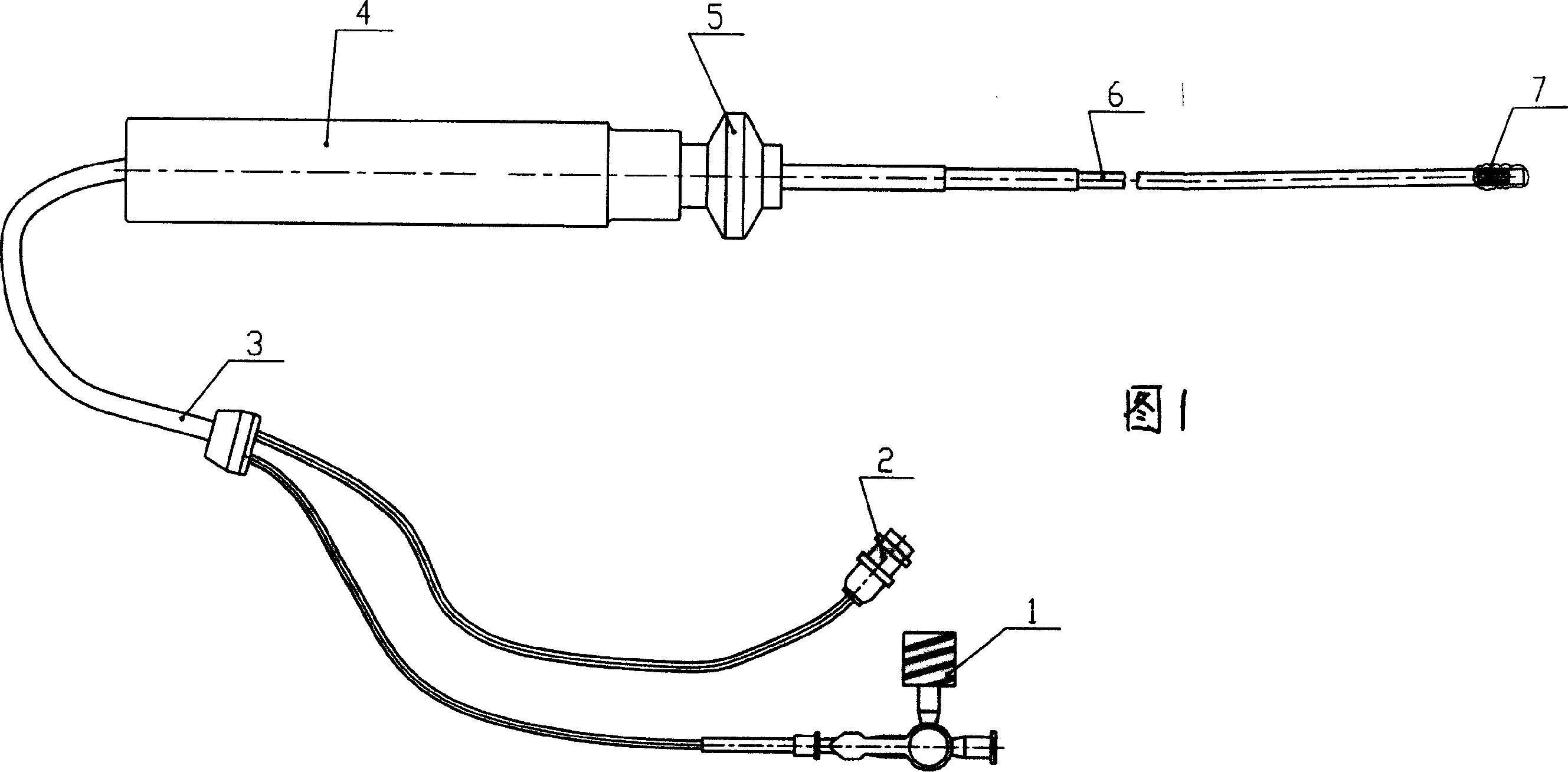
[0028] The overall dimensions of the detection catheter are similar to that of radiofrequency ablation leads. The outer diameter of the catheter is 6F. With the help of the vascular sheath, it can enter the left ventricle, aortic arch, thoracoabdominal aorta, etc. through the femoral artery and radial artery. It can enter the vena cava, right heart system and pulmonary artery through the femoral vein. Enter the stomach, esophagus, colon, or uterus directly or with the help of an endoscope; enter the thoracic and abdominal cavities with the help of body laparoscopy (such as with a laparoscope) to enter the abdominal cavity.

[0029] The controllable bending principle of the detection catheter is similar to that of the radiofrequency ablation lead. The hardness parameters are obtained by applying stress to the examined tissue, or by means of the contracti...

Embodiment 2

[0032] Example 2, the basic operation process of the method of acquiring tissue hardness information by interventional ultrasound

[0033] The catheter is sent into the body in the form of an ordinary interventional catheter. The catheter head is in contact with the water bladder and the surface of the tissue with different strengths along the vertical direction of the tissue to be tested. The pressure on the tissue is transmitted to the pressure sensor through the water bladder liquid to obtain a real-time pressure curve. After the water bladder touches the pressure, a certain deformation occurs in the direction of the long axis of the catheter. The deformation is detected by high-resolution ultrasound and displayed and recorded synchronously with the pressure and ECG curves on the same screen in M-mode ultrasound mode. Using the computer software provided by this solution, the hardness constant K value can be obtained by synchronously measuring the pressure applied to the tis...

Embodiment 3

[0035] Example 3 Method for Acquiring Catheter Ultrasonic Hardness Information

[0036] The method includes the following steps:

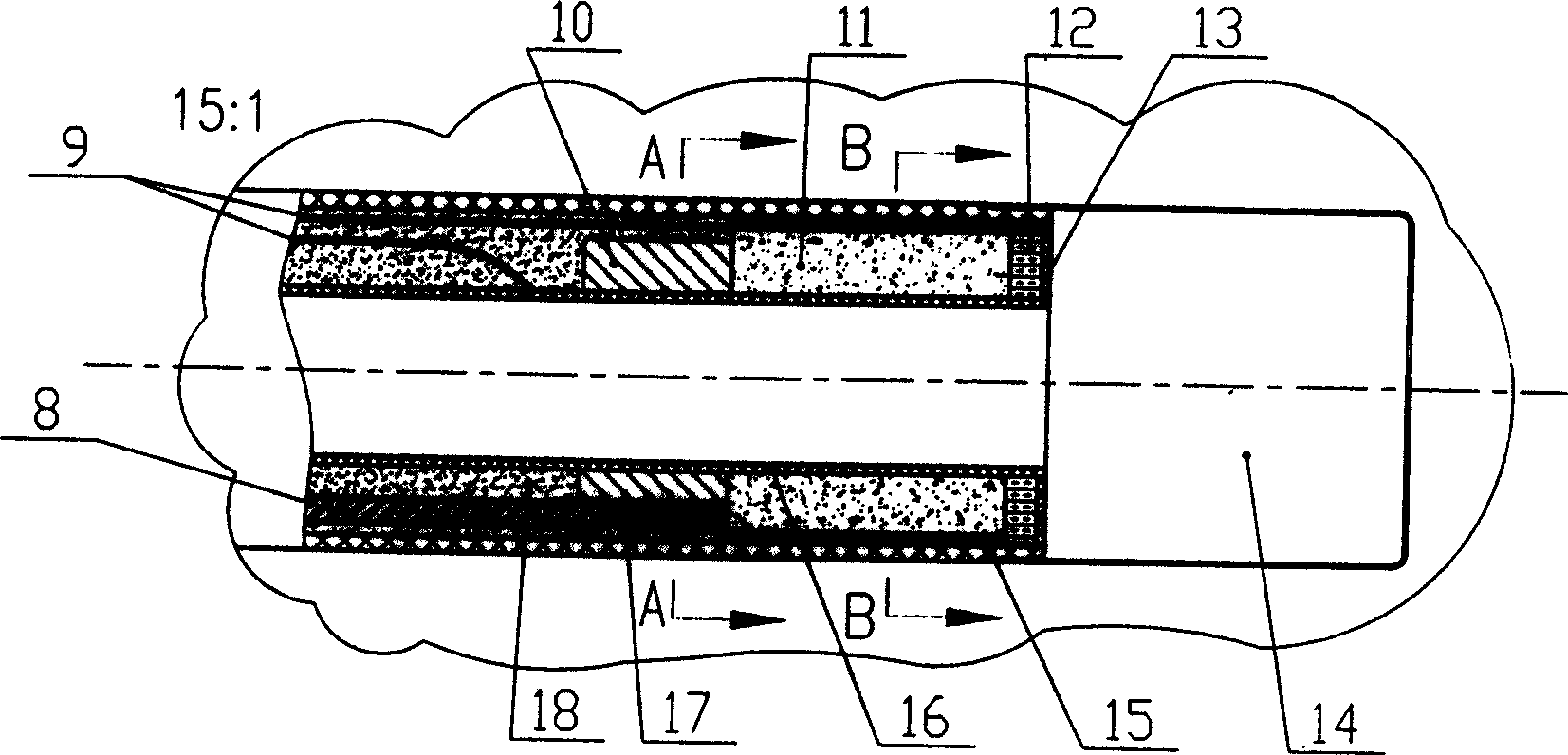
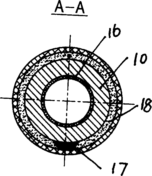
[0037] a. Establish or set up the ultrasonic transducer and pressure sensor structure carried by the interventional catheter;

[0038] b. The pressure is applied to the examined tissue through the interventional catheter, and the applied force is continuously recorded by the pressure sensor in real time; at the same time, the ultrasonic transducer receives the change of the echo signal in the process of the pressure of the examined tissue in real time and continuously, and passes the M-mode The echocardiographic mode shows images of changes in tissue thickness.

[0039] c. Establish a superimposition or comparison graphic between the pressure signal curve and the image showing tissue thickness changes in the M-mode echocardiography mode; select a good image freeze frame for detection of pressure and deformation thickness to be displayed synchronou...


PUM


Abstract

The invention relates to one kind of tissue hardness acquisition method by intervenient ultrasound and comprises building ultrasonic transducer supported by intervenient duct and pressure sensor structure. By intervenient duct operated out of body, forcing pressure to detected tissue through aqueous capsule, recordings continually pressure and the echo signal variety received by ultrasonic transducer, and displaying variety image of tissue thickness by M type ultrasound cardiogram pattern; measuring the change values of pressure and thickness; charactering tissue hardness by pressure value and change value of thickness obtained, and computing elastic modulus. Intervenient ultrasound hardness detector mentioned comprises ultrasound hardness detecting duct and host computer, liquid sac is fixed on the head end of duct, and the ultrasonic transducer and pressure sensor following fixed; power line and signal line in duct connect to main computer. By this invention people can obtain directly tissue hardness for passive deforming ability, elastic modulus and other basic physical quantity, which brings direct and reliable information to scientific study and detecting tissue hardness in clinic.

Description

technical field [0001] The invention relates to a method for acquiring physical parameters of a human body, an ultrasonic diagnostic instrument, and at the same time relates to an intervention minimally invasive information acquisition technology. It is used to obtain the hardness value of myocardium, great blood vessels and other deep internal organs of the human body. It is a method to obtain and analyze the tissue stress-strain relationship in the deep part of human or animal living body by catheter intervention technology, so as to directly obtain the elastic parameters and properties of human tissue. The information obtained is useful in assisting medical research and clinical diagnosis. Background technique: [0002] The hardness of living tissue is an important indicator to characterize whether the tissue is diseased, and it plays an important role in judging the changes in the hardness of myocardial, blood vessel, and visceral tissue. Myocardial ischemia, old myoca...

Claims


Application Information

Patent Timeline
Patent Type & Authority Applications(China)
IPC IPC(8): A61B8/00A61B8/12
Inventor 黄晶邓昌明郑永平凌智瑜刘地川邓辉胜
Owner 黄晶
Why Eureka
Try Eureka